 Français
 English
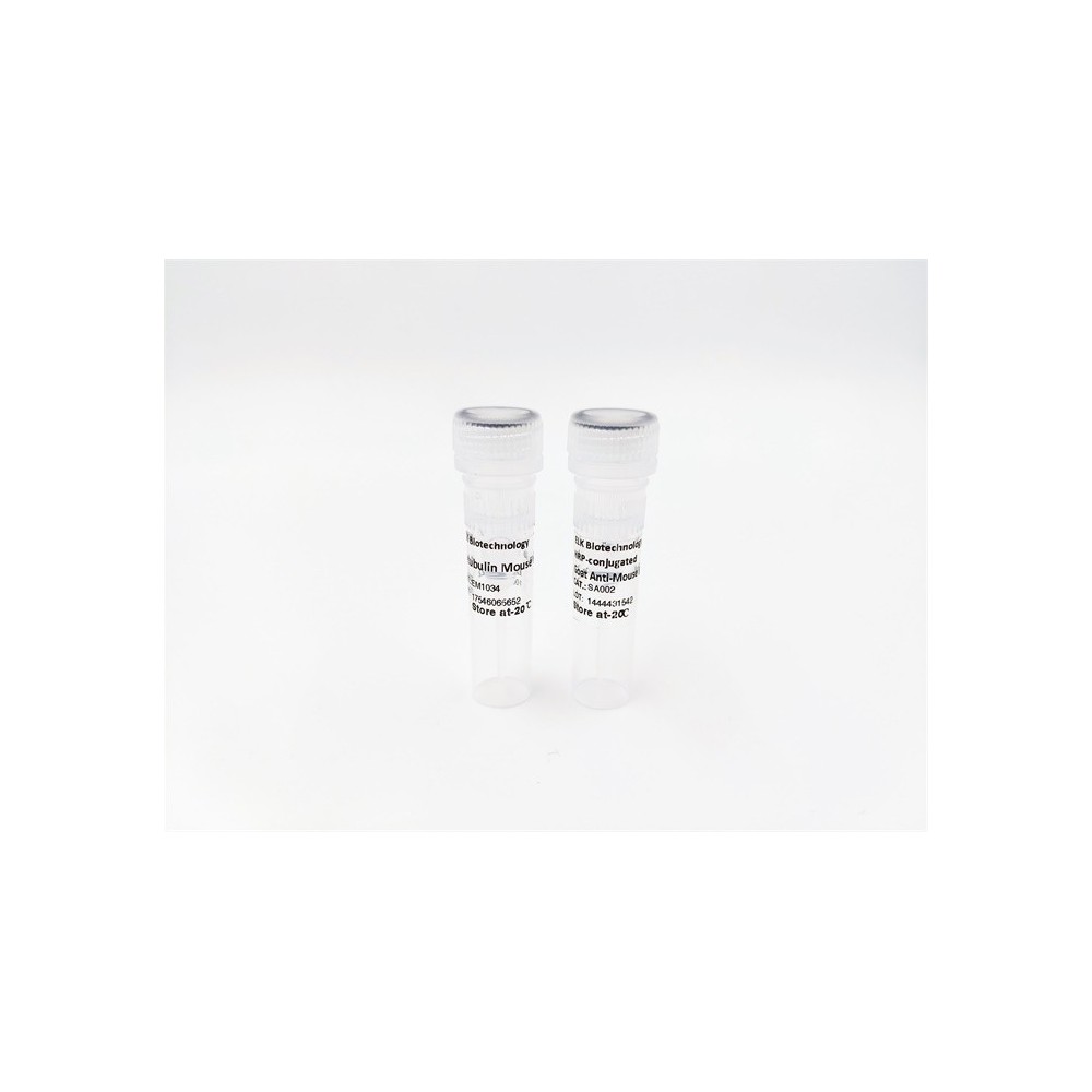

| ELK.No | ES2052 |
| Product name | Crk II rabbit pAb |
| Reactivity | Human;Mouse;Rat;Monkey |
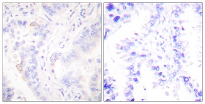
| Applications | WB;IHC;IF;ELISA |
| Other name | CRK; Adapter molecule crk; Proto-oncogene c-Crk; p38 |
| Size | 50μL |
| Unit price ($) | 148 |
| Human gene ID | 1398 |
| Human Swiss-Prot | P46108 |
| Source | Rabbit |
| Isotype | IgG |
| Target | Crk II |
| Fields | >>MAPK signaling pathway;>>ErbB signaling pathway;>>Rap1 signaling pathway;>>Chemokine signaling pathway;>>Focal adhesion;>>Fc gamma R-mediated phagocytosis;>>Neurotrophin signaling pathway;>>Regulation of actin cytoskeleton;>>Insulin signaling pathway;>>Growth hormone synthesis, secretion and action;>>Bacterial invasion of epithelial cells;>>Shigellosis;>>Yersinia infection;>>Human cytomegalovirus infection;>>Human immunodeficiency virus 1 infection;>>Pathways in cancer;>>MicroRNAs in cancer;>>Renal cell carcinoma;>>Chronic myeloid leukemia |
| Gene name | CRK |
| Protein name | Adapter molecule crk |
| Human gene link | View Human Gene |
| Human Swiss link | View Human Swiss-Prot |
| Mouse gene ID | 12928 |
| Mouse gene link | View Mouse Gene |
| Mouse Swiss-Prot | Q64010 |
| Mouse Swiss link | View Mouse Swiss-Prot |
| Rat gene ID | 54245 |
| Rat gene link | View Rat Gene |
| Rat Swiss-Prot | Q63768 |
| Rat Swiss link | View Rat Swiss-Prot |
| Immunogen | The antiserum was produced against synthesized peptide derived from human CrkII. AA range:187-236 |
| Specificity | Crk II Polyclonal Antibody detects endogenous levels of Crk II protein. |
| Formulation | Liquid in PBS containing 50% glycerol, 0.5% BSA and 0.02% sodium azide. |
| Clonality | Polyclonal |
| Dilution | Western Blot: 1/500 - 1/2000. Immunohistochemistry: 1/100 - 1/300. Immunofluorescence: 1/200 - 1/1000. ELISA: 1/5000. Not yet tested in other applications. |
| Purification | The antibody was affinity-purified from rabbit antiserum by affinity-chromatography using epitope-specific immunogen. |
| Concentration | 1 mg/ml |
| Storage stability | -20°C/1 year |
| Molecular Weight (Da) | |
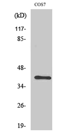
| Observed band (KD) | 40kD |
| Background | This gene encodes a member of an adapter protein family that binds to several tyrosine-phosphorylated proteins. The product of this gene has several SH2 and SH3 domains (src-homology domains) and is involved in several signaling pathways, recruiting cytoplasmic proteins in the vicinity of tyrosine kinase through SH2-phosphotyrosine interaction. The N-terminal SH2 domain of this protein functions as a positive regulator of transformation whereas the C-terminal SH3 domain functions as a negative regulator of transformation. Two alternative transcripts encoding different isoforms with distinct biological activity have been described. [provided by RefSeq, Jul 2008], |
| Function | domain:The C-terminal SH3 domain function as a negative modulator for transformation and the N-terminal SH3 domain appears to function as a positive regulator for transformation.,domain:The SH2 domain mediates interaction with SHB.,function:The Crk-I and Crk-II forms differ in their biological activities. Crk-II has less transforming activity than Crk-I. Crk-II mediates attachment-induced MAPK8 activation, membrane ruffling and cell motility in a Rac-dependent manner. Involved in phagocytosis of apoptotic cells and cell motility via its interaction with DOCK1 and DOCK4.,PTM:Phosphorylated on Tyr-221 upon cell adhesion. Results in the negative regulation of the association with SH2- and SH3-binding partners, possibly by the formation of an intramolecular interaction of phosphorylated Tyr-221 with the SH2 domain. This leads finally to the down-regulation of the Crk signaling pathway.,PTM:P |
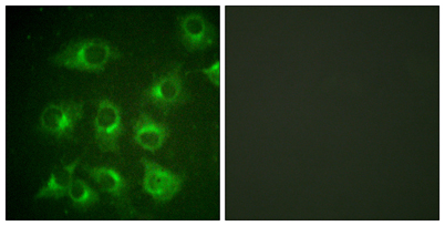
| Subcellular location | Cytoplasm . Cell membrane . Translocated to the plasma membrane upon cell adhesion. . |
| Expression | Embryonic lung,Epithelium,Eye,Lung,Placenta, |

Use collapsible tabs for more detailed information that will help customers make a purchasing decision.
Ex: Shipping and return policies, size guides, and other common questions.