 Français
 English
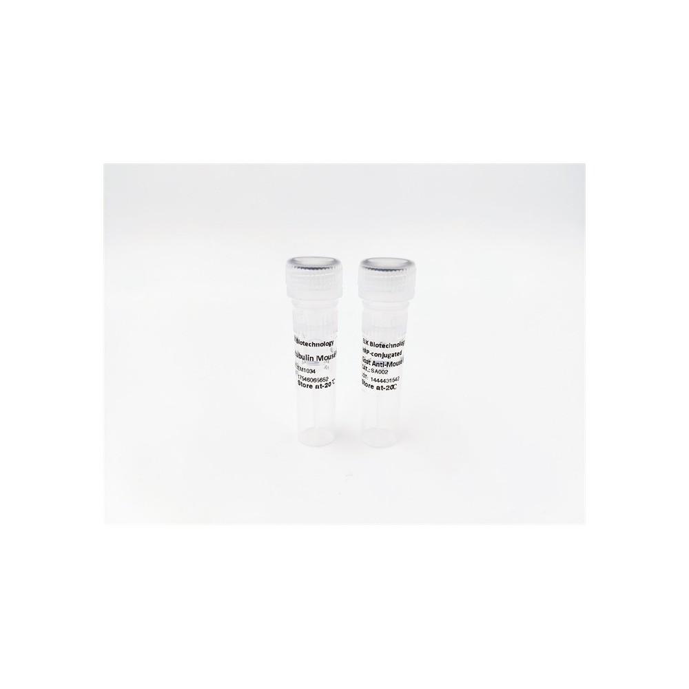

| ELK.No | ES2380 |
| Product name | Fusin rabbit pAb |
| Reactivity | Human;Mouse;Rat |
| Applications | WB;IF;ELISA |
| Other name | CXCR4; C-X-C chemokine receptor type 4; CXC-R4; CXCR-4; FB22; Fusin; HM89; LCR1; Leukocyte-derived seven transmembrane domain receptor; LESTR; NPYRL; Stromal cell-derived factor 1 receptor; SDF-1 receptor; CD antigen CD184 |
| Size | 100μL |
| Unit price ($) | 248 |
| Human gene ID | 7852 |
| Human Swiss-Prot | P61073 |
| Source | Rabbit |
| Isotype | IgG |
| Target | CXCR4 |
| Fields | >>Viral life cycle - HIV-1;>>Calcium signaling pathway;>>Cytokine-cytokine receptor interaction;>>Viral protein interaction with cytokine and cytokine receptor;>>Chemokine signaling pathway;>>Endocytosis;>>Axon guidance;>>Leukocyte transendothelial migration;>>Intestinal immune network for IgA production;>>Regulation of actin cytoskeleton;>>Human cytomegalovirus infection;>>Human immunodeficiency virus 1 infection;>>Pathways in cancer |
| Gene name | CXCR4 |
| Protein name | C-X-C chemokine receptor type 4 |
| Human gene link | View Human Gene |
| Human Swiss link | View Human Swiss-Prot |
| Mouse gene ID | 12767 |
| Mouse gene link | View Mouse Gene |
| Mouse Swiss-Prot | P70658 |
| Mouse Swiss link | View Mouse Swiss-Prot |
| Rat gene ID | |
| Rat gene link | |
| Rat Swiss-Prot | O08565 |
| Rat Swiss link | View Rat Swiss-Prot |
| Immunogen | The antiserum was produced against synthesized peptide derived from human CXCR4. AA range:300-349 |
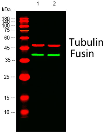
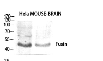
| Specificity | Fusin Polyclonal Antibody detects endogenous levels of Fusin protein. |
| Formulation | Liquid in PBS containing 50% glycerol, 0.5% BSA and 0.02% sodium azide. |
| Clonality | Polyclonal |
| Dilution | Western Blot: 1/500 - 1/2000. Immunofluorescence: 1/200 - 1/1000. ELISA: 1/40000. Not yet tested in other applications. |
| Purification | The antibody was affinity-purified from rabbit antiserum by affinity-chromatography using epitope-specific immunogen. |
| Concentration | 1 mg/ml |
| Storage stability | -20°C/1 year |
| Molecular Weight (Da) | |
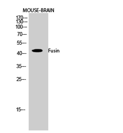
| Observed band (KD) | 36kD |
| Background | C-X-C motif chemokine receptor 4(CXCR4) Homo sapiens This gene encodes a CXC chemokine receptor specific for stromal cell-derived factor-1. The protein has 7 transmembrane regions and is located on the cell surface. It acts with the CD4 protein to support HIV entry into cells and is also highly expressed in breast cancer cells. Mutations in this gene have been associated with WHIM (warts, hypogammaglobulinemia, infections, and myelokathexis) syndrome. Alternate transcriptional splice variants, encoding different isoforms, have been characterized. [provided by RefSeq, Jul 2008], |
| Function | alternative products:Additional isoforms seem to exist,caution:Was originally (PubMed:8329116 and PubMed:8234909) thought to be a receptor for neuropeptide Y type 3 (NPY3R) (NPY3-R).,disease:Defects in CXCR4 are a cause of WHIM syndrome [MIM:193670]; also called warts, hypogammaglobulinemia, infections, and myelokathexis. WHIM syndrome is an immunodeficiency disease characterized by neutropenia, hypogammaglobulinemia and extensive human papillomavirus (HPV) infection. Despite the peripheral neutropenia, bone marrow aspirates from affected individuals contain abundant mature myeloid cells, a condition termed myelokathexis.,domain:The amino-terminus is critical for ligand binding. Residues in all four extracellular regions contribute to HIV-1 coreceptor activity.,function:Receptor for the C-X-C chemokine CXCL12/SDF-1. Transduces a signal by increasing the intracellular calcium ions level. |
| Subcellular location | Cell membrane ; Multi-pass membrane protein . Cell junction. Early endosome. Late endosome. Lysosome. In unstimulated cells, diffuse pattern on plasma membrane. On agonist stimulation, colocalizes with ITCH at the plasma membrane where it becomes ubiquitinated. In the presence of antigen, distributes to the immunological synapse forming at the T-cell-APC contact area, where it localizes at the peripheral and distal supramolecular activation cluster (SMAC). |
| Expression | Expressed in numerous tissues, such as peripheral blood leukocytes, spleen, thymus, spinal cord, heart, placenta, lung, liver, skeletal muscle, kidney, pancreas, cerebellum, cerebral cortex and medulla (in microglia as well as in astrocytes), brain microvascular, coronary artery and umbilical cord endothelial cells. Isoform 1 is predominant in all tissues tested. |

Use collapsible tabs for more detailed information that will help customers make a purchasing decision.
Ex: Shipping and return policies, size guides, and other common questions.