 Français
 English
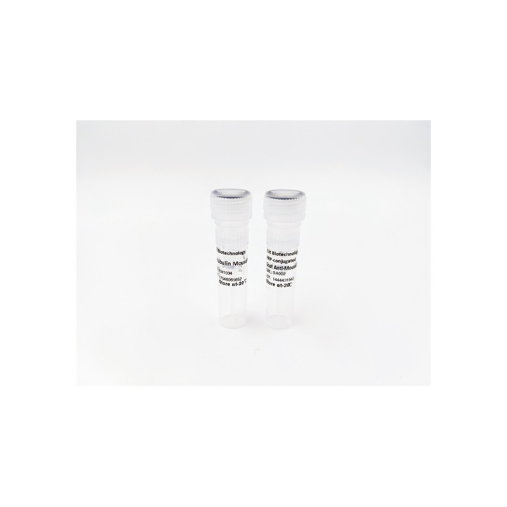

| ELK.No | ES2586 |
| Product name | HURP rabbit pAb |
| Reactivity | Human;Rat;Mouse; |
| Applications | WB;ELISA |
| Other name | DLGAP5; DLG7; KIAA0008; Disks large-associated protein 5; DAP-5; Discs large homolog 7; Disks large-associated protein DLG7; Hepatoma up-regulated protein; HURP |
| Size | 50μL |
| Unit price ($) | 148 |
| Human gene ID | 9787 |
| Human Swiss-Prot | Q15398 |
| Source | Rabbit |
| Isotype | IgG |
| Target | HURP |
| Fields | |
| Gene name | DLGAP5 |
| Protein name | Disks large-associated protein 5 |
| Human gene link | View Human Gene |
| Human Swiss link | View Human Swiss-Prot |
| Mouse gene ID | |
| Mouse gene link | |
| Mouse Swiss-Prot | Q8K4R9 |
| Mouse Swiss link | View Mouse Swiss-Prot |
| Rat gene ID | |
| Rat gene link | |
| Rat Swiss-Prot | |
| Rat Swiss link | |
| Immunogen | The antiserum was produced against synthesized peptide derived from human DLGAP5. AA range:791-840 |
| Specificity | HURP Polyclonal Antibody detects endogenous levels of HURP protein. |
| Formulation | Liquid in PBS containing 50% glycerol, 0.5% BSA and 0.02% sodium azide. |
| Clonality | Polyclonal |
| Dilution | Western Blot: 1/500 - 1/2000. ELISA: 1/40000. Not yet tested in other applications. |
| Purification | The antibody was affinity-purified from rabbit antiserum by affinity-chromatography using epitope-specific immunogen. |
| Concentration | 1 mg/ml |
| Storage stability | -20°C/1 year |
| Molecular Weight (Da) | |
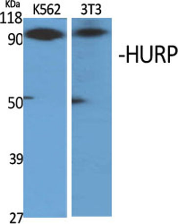
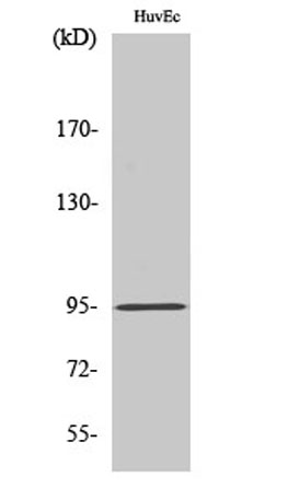
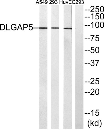
| Observed band (KD) | 95kD |
| Background | developmental stage:Elevated levels of expression detected in the G2/M phase of synchronized cultures of HeLa cells.,function:Potential cell cycle regulator that may play a role in carcinogenesis of cancer cells. Mitotic phosphoprotein regulated by the ubiquitin-proteasome pathway. Key regulator of adherens junction integrity and differentiation that may be involved in CDH1-mediated adhesion and signaling in epithelial cells.,PTM:Phosphorylated upon DNA damage, probably by ATM or ATR. Decreased phosphorylation levels are associated with the differentiation of intestinal epithelial cells.,PTM:Ubiquitinated, leading to its degradation.,similarity:Belongs to the SAPAP family.,subcellular location:Localizes to the spindle poles in mitotic cells. Colocalizes with CDH1 at sites of cell-cell contact in intestinal epithelial cells.,subunit:Interacts with CDC2. Interacts with the C-terminal proline-rich region of FBXO7. Recruited by FBXO7 to a SCF (SKP1-CUL1-F-box) protein complex in a CDC2/Cyclin B-phosphorylation dependent manner. Interacts with CDH1.,tissue specificity:Abundantly expressed in fetal liver. Expressed at lower levels in bone marrow, testis, colon, and placenta., |
| Function | developmental stage:Elevated levels of expression detected in the G2/M phase of synchronized cultures of HeLa cells.,function:Potential cell cycle regulator that may play a role in carcinogenesis of cancer cells. Mitotic phosphoprotein regulated by the ubiquitin-proteasome pathway. Key regulator of adherens junction integrity and differentiation that may be involved in CDH1-mediated adhesion and signaling in epithelial cells.,PTM:Phosphorylated upon DNA damage, probably by ATM or ATR. Decreased phosphorylation levels are associated with the differentiation of intestinal epithelial cells.,PTM:Ubiquitinated, leading to its degradation.,similarity:Belongs to the SAPAP family.,subcellular location:Localizes to the spindle poles in mitotic cells. Colocalizes with CDH1 at sites of cell-cell contact in intestinal epithelial cells.,subunit:Interacts with CDC2. Interacts with the C-terminal proli |
| Subcellular location | Nucleus. Cytoplasm. Cytoplasm, cytoskeleton, spindle. Localizes to the spindle in mitotic cells. Colocalizes with CDH1 at sites of cell-cell contact in intestinal epithelial cells. |
| Expression | Abundantly expressed in fetal liver. Expressed at lower levels in bone marrow, testis, colon, and placenta. |

Use collapsible tabs for more detailed information that will help customers make a purchasing decision.
Ex: Shipping and return policies, size guides, and other common questions.