 Français
 English

| ELK.No | ES2628 |
| Product name | Integrin α3 rabbit pAb |
| Reactivity | Human;Mouse;Rat |
| Applications | WB;ELISA |
| Other name | ITGA3; MSK18; Integrin alpha-3; CD49 antigen-like family member C; FRP-2; Galactoprotein B3; GAPB3; VLA-3 subunit alpha; CD antigen CD49c |
| Size | 100μL |
| Unit price ($) | 248 |
| Human gene ID | 3675 |
| Human Swiss-Prot | P26006 |
| Source | Rabbit |
| Isotype | IgG |
| Target | Integrin α3 |
| Fields | >>PI3K-Akt signaling pathway;>>Focal adhesion;>>ECM-receptor interaction;>>Hematopoietic cell lineage;>>Regulation of actin cytoskeleton;>>Human papillomavirus infection;>>Pathways in cancer;>>Small cell lung cancer;>>Hypertrophic cardiomyopathy;>>Arrhythmogenic right ventricular cardiomyopathy;>>Dilated cardiomyopathy |
| Gene name | ITGA3 |
| Protein name | Integrin alpha-3 |
| Human gene link | View Human Gene |
| Human Swiss link | View Human Swiss-Prot |
| Mouse gene ID | 16400 |
| Mouse gene link | View Mouse Gene |
| Mouse Swiss-Prot | Q62470 |
| Mouse Swiss link | View Mouse Swiss-Prot |
| Rat gene ID | |
| Rat gene link | |
| Rat Swiss-Prot | |
| Rat Swiss link | |
| Immunogen | The antiserum was produced against synthesized peptide derived from human Integrin alpha3. AA range:482-531 |
| Specificity | Integrin α3 Polyclonal Antibody detects endogenous levels of Integrin α3 protein. |
| Formulation | Liquid in PBS containing 50% glycerol, 0.5% BSA and 0.02% sodium azide. |
| Clonality | Polyclonal |
| Dilution | Western Blot: 1/500 - 1/2000. ELISA: 1/40000. Not yet tested in other applications. |
| Purification | The antibody was affinity-purified from rabbit antiserum by affinity-chromatography using epitope-specific immunogen. |
| Concentration | 1 mg/ml |
| Storage stability | -20°C/1 year |
| Molecular Weight (Da) | |
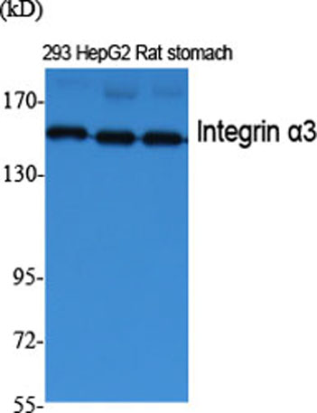
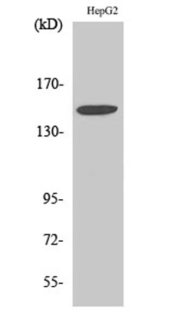
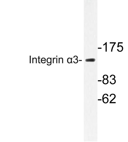
| Observed band (KD) | 150kD |
| Background | integrin subunit alpha 3(ITGA3) Homo sapiens The gene encodes a member of the integrin alpha chain family of proteins. Integrins are heterodimeric integral membrane proteins composed of an alpha chain and a beta chain that function as cell surface adhesion molecules. The encoded preproprotein is proteolytically processed to generate light and heavy chains that comprise the alpha 3 subunit. This subunit joins with a beta 1 subunit to form an integrin that interacts with extracellular matrix proteins including members of the laminin family. Expression of this gene may be correlated with breast cancer metastasis. [provided by RefSeq, Oct 2015], |
| Function | function:Integrin alpha-3/beta-1 is a receptor for fibronectin, laminin, collagen, epiligrin, thrombospondin and CSPG4. Alpha-3/beta-1 may mediate with LGALS3 the stimulation by CSPG4 of endothelial cells migration.,PTM:Isoform alpha-3A, but not isoform alpha-3B, is phosphorylated on serine residues. Phosphorylation increases after phorbol 12-myristate 13-acetate stimulation. Isoform alpha-3A is phosphorylated on Tyr-1051.,similarity:Belongs to the integrin alpha chain family.,similarity:Contains 7 FG-GAP repeats.,subunit:Heterodimer of an alpha and a beta subunit. The alpha subunit is composed of an heavy and a light chain linked by a disulfide bond. Alpha-3 associates with beta-1. Interacts with HPS5.,tissue specificity:Isoform alpha-3A is widely expressed. Isoform alpha-3B is expressed in brain and heart. In brain, both isoforms are exclusively expressed on vascular smooth muscle cell |
| Subcellular location | Cell membrane ; Single-pass type I membrane protein . Cell membrane ; Lipid-anchor . Cell projection, invadopodium membrane ; Single-pass type I membrane protein . Cell projection, filopodium membrane ; Single-pass type I membrane protein . Enriched preferentially at invadopodia, cell membrane protrusions that correspond to sites of cell invasion, in a collagen-dependent manner. . |
| Expression | Isoform 1 is widely expressed. Isoform 2 is expressed in brain and heart. In brain, both isoforms are exclusively expressed on vascular smooth muscle cells, whereas in heart isoform 1 is strongly expressed on vascular smooth muscle cells, isoform 2 is detected only on endothelial vein cells. |

Use collapsible tabs for more detailed information that will help customers make a purchasing decision.
Ex: Shipping and return policies, size guides, and other common questions.