 Français
 English

| ELK.No | ES2642 |
| Product name | IRAK-2 rabbit pAb |
| Reactivity | Human;Rat;Mouse; |
| Applications | WB;IHC |
| Other name | IRAK2; Interleukin-1 receptor-associated kinase-like 2; IRAK-2 |
| Size | 100μL |
| Unit price ($) | 248 |
| Human gene ID | 3656 |
| Human Swiss-Prot | O43187 |
| Source | Rabbit |
| Isotype | IgG |
| Target | IRAK-2 |
| Fields | >>Neurotrophin signaling pathway;>>Tuberculosis |
| Gene name | IRAK2 |
| Protein name | Interleukin-1 receptor-associated kinase-like 2 |
| Human gene link | View Human Gene |
| Human Swiss link | View Human Swiss-Prot |
| Mouse gene ID | |
| Mouse gene link | |
| Mouse Swiss-Prot | Q8CFA1 |
| Mouse Swiss link | View Mouse Swiss-Prot |
| Rat gene ID | |
| Rat gene link | |
| Rat Swiss-Prot | |
| Rat Swiss link | |
| Immunogen | Synthesized peptide derived from the Internal region of human IRAK-2. |
| Specificity | IRAK-2 Polyclonal Antibody detects endogenous levels of IRAK-2 protein. |
| Formulation | Liquid in PBS containing 50% glycerol, 0.5% BSA and 0.02% sodium azide. |
| Clonality | Polyclonal |
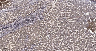
| Dilution | WB 1:500-2000;IHC-p 1:50-300 |
| Purification | The antibody was affinity-purified from rabbit antiserum by affinity-chromatography using epitope-specific immunogen. |
| Concentration | 1 mg/ml |
| Storage stability | -20°C/1 year |
| Molecular Weight (Da) | |
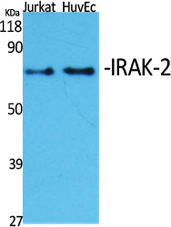
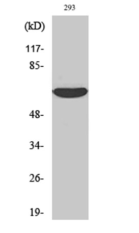
| Observed band (KD) | 70kD |
| Background | IRAK2 encodes the interleukin-1 receptor-associated kinase 2, one of two putative serine/threonine kinases that become associated with the interleukin-1 receptor (IL1R) upon stimulation. IRAK2 is reported to participate in the IL1-induced upregulation of NF-kappaB. [provided by RefSeq, Jul 2008], |
| Function | caution:Asn-335 is present instead of the conserved Asp which is expected to be an active site residue. This enzyme has been shown to be catalytically inactive.,domain:The protein kinase domain is predicted to be catalytically inactive.,function:Binds to the IL-1 type I receptor following IL-1 engagement, triggering intracellular signaling cascades leading to transcriptional up-regulation and mRNA stabilization.,similarity:Belongs to the protein kinase superfamily. TKL Ser/Thr protein kinase family. Pelle subfamily.,similarity:Contains 1 death domain.,similarity:Contains 1 protein kinase domain.,subunit:Interacts with MYD88. IL-1 stimulation leads to the formation of a signaling complex which dissociates from the IL-1 receptor following the binding of PELI1.,tissue specificity:Expressed in spleen, thymus, prostate, lung, liver, skeletal muscle, kidney, pancreas and peripheral blood leuko |
| Subcellular location | nucleus,cytoplasm,cytosol,plasma membrane,endosome membrane, |
| Expression | Expressed in spleen, thymus, prostate, lung, liver, skeletal muscle, kidney, pancreas and peripheral blood leukocytes. |

Use collapsible tabs for more detailed information that will help customers make a purchasing decision.
Ex: Shipping and return policies, size guides, and other common questions.