 Français
 English
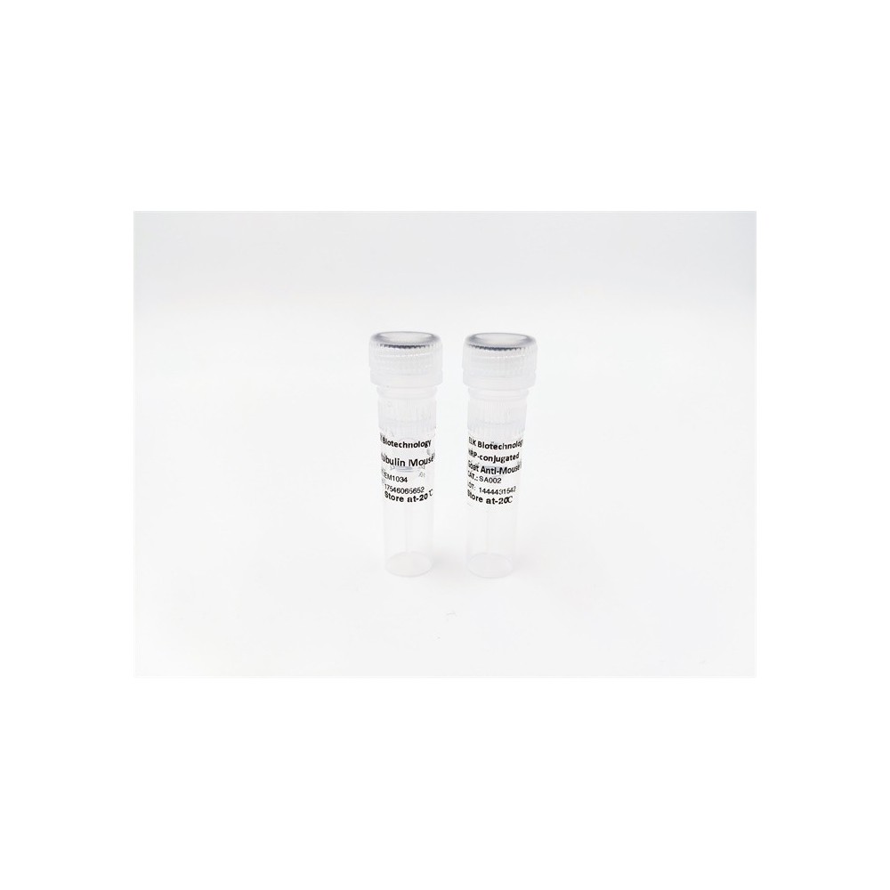

| ELK.No | ES2739 |
| Product name | MADD rabbit pAb |
| Reactivity | Human;Mouse;Rat |
| Applications | WB;IHC;IF;ELISA |
| Other name | MADD; DENN; IG20; KIAA0358; MAP kinase-activating death domain protein; Differentially expressed in normal and neoplastic cells; Insulinoma glucagonoma clone 20; Rab3 GDP/GTP exchange factor |
| Size | 100μL |
| Unit price ($) | 248 |
| Human gene ID | 8567 |
| Human Swiss-Prot | Q8WXG6 |
| Source | Rabbit |
| Isotype | IgG |
| Target | MADD |
| Fields | |
| Gene name | MADD |
| Protein name | MAP kinase-activating death domain protein |
| Human gene link | View Human Gene |
| Human Swiss link | View Human Swiss-Prot |
| Mouse gene ID | 228355 |
| Mouse gene link | View Mouse Gene |
| Mouse Swiss-Prot | Q80U28 |
| Mouse Swiss link | View Mouse Swiss-Prot |
| Rat gene ID | 94193 |
| Rat gene link | View Rat Gene |
| Rat Swiss-Prot | O08873 |
| Rat Swiss link | View Rat Swiss-Prot |
| Immunogen | The antiserum was produced against synthesized peptide derived from human MADD. AA range:751-800 |
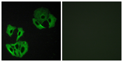
| Specificity | MADD Polyclonal Antibody detects endogenous levels of MADD protein. |
| Formulation | Liquid in PBS containing 50% glycerol, 0.5% BSA and 0.02% sodium azide. |
| Clonality | Polyclonal |
| Dilution | Western Blot: 1/500 - 1/2000. Immunohistochemistry: 1/100 - 1/300. Immunofluorescence: 1/200 - 1/1000. ELISA: 1/5000. Not yet tested in other applications. |
| Purification | The antibody was affinity-purified from rabbit antiserum by affinity-chromatography using epitope-specific immunogen. |
| Concentration | 1 mg/ml |
| Storage stability | -20°C/1 year |
| Molecular Weight (Da) | |
| Observed band (KD) | 183kD |
| Background | Tumor necrosis factor alpha (TNF-alpha) is a signaling molecule that interacts with one of two receptors on cells targeted for apoptosis. The apoptotic signal is transduced inside these cells by cytoplasmic adaptor proteins. The protein encoded by this gene is a death domain-containing adaptor protein that interacts with the death domain of TNF-alpha receptor 1 to activate mitogen-activated protein kinase (MAPK) and propagate the apoptotic signal. It is membrane-bound and expressed at a higher level in neoplastic cells than in normal cells. Several transcript variants encoding different isoforms have been described for this gene. [provided by RefSeq, Jul 2008], |
| Function | caution:The sequence shown here is derived from an Ensembl automatic analysis pipeline and should be considered as preliminary data.,function:Plays a significant role in regulating cell proliferation, survival and death through alternative mRNA splicing. Isoform 5 shows increased cell proliferation and isoform 2 shows decreased. Converts GDP-bound inactive form of RAB3A, RAB3C and RAB3D to the GTP-bound active forms. Component of the TNFRSF1A signaling complex: MADD links TNFRSF1A with MAP kinase activation. Plays an important regulatory role in physiological cell death (TNF-alpha-induced, caspase-mediated apoptosis); isoform 1 is susceptible to inducing apoptosis, isoform 5 is resistant and isoform 3 and isoform 4 have no effect.,miscellaneous:Overexpression of MADD activates the mitogen-activated protein (MAP) kinase extracellular signal-regulated kinase (ERK). Expression of the MADD d |
| Subcellular location | Cell membrane . Cytoplasm . Cell projection, axon . |
| Expression | Expressed in testis, ovary, brain and heart (PubMed:8988362). Expressed in spleen, thymus, prostate, testis, ovary, small instestine and colon (PubMed:9115275). Expressed in liver (PubMed:9796103). ; [Isoform 1]: Not detected in the brain, breast, kidney, lung, ovary, pancreas, testis, uterus, stomach and thyroid. ; [Isoform 2]: Expressed in the brain, breast, kidney, lung, ovary, pancreas, testis, uterus, stomach and thyroid. ; [Isoform 3]: Expressed in the brain, breast, kidney, lung, ovary, pancreas, testis, uterus, stomach and thyroid. ; [Isoform 4]: Expressed in the brain, breast, kidney, lung, ovary, pancreas, testis, uterus, stomach and thyroid. ; [Isoform 5]: Expressed in the brain, breast, kidney, lung, ovary, pancreas, testis, uterus, stomach and thyroid. ; [Isoform 6]: Not detec |
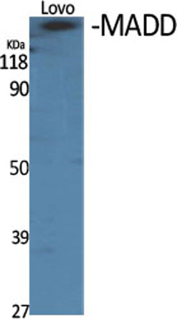
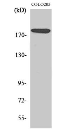

Use collapsible tabs for more detailed information that will help customers make a purchasing decision.
Ex: Shipping and return policies, size guides, and other common questions.