 Français
 English

| ELK.No | ES3146 |
| Product name | PAKα rabbit pAb |
| Reactivity | Human;Mouse;Rat |
| Applications | WB;IHC;IF;ELISA |
| Other name | PAK1; Serine/threonine-protein kinase PAK 1; Alpha-PAK; p21-activated kinase 1; PAK-1; p65-PAK |
| Size | 100μL |
| Unit price ($) | 248 |
| Human gene ID | 5058 |
| Human Swiss-Prot | Q13153 |
| Source | Rabbit |
| Isotype | IgG |
| Target | PAK1 |
| Fields | >>MAPK signaling pathway;>>ErbB signaling pathway;>>Ras signaling pathway;>>cAMP signaling pathway;>>Chemokine signaling pathway;>>Axon guidance;>>Hippo signaling pathway - multiple species;>>Focal adhesion;>>C-type lectin receptor signaling pathway;>>Natural killer cell mediated cytotoxicity;>>T cell receptor signaling pathway;>>Fc gamma R-mediated phagocytosis;>>Regulation of actin cytoskeleton;>>Epithelial cell signaling in Helicobacter pylori infection;>>Pathogenic Escherichia coli infection;>>Salmonella infection;>>Human immunodeficiency virus 1 infection;>>Proteoglycans in cancer;>>Renal cell carcinoma |
| Gene name | PAK1 |
| Protein name | Serine/threonine-protein kinase PAK 1 |
| Human gene link | View Human Gene |
| Human Swiss link | View Human Swiss-Prot |
| Mouse gene ID | |
| Mouse gene link | |
| Mouse Swiss-Prot | O88643 |
| Mouse Swiss link | View Mouse Swiss-Prot |
| Rat gene ID | 29431 |
| Rat gene link | View Rat Gene |
| Rat Swiss-Prot | P35465 |
| Rat Swiss link | View Rat Swiss-Prot |
| Immunogen | The antiserum was produced against synthesized peptide derived from human PAK1. AA range:178-227 |
| Specificity | PAKα Polyclonal Antibody detects endogenous levels of PAKα protein. |
| Formulation | Liquid in PBS containing 50% glycerol, 0.5% BSA and 0.02% sodium azide. |
| Clonality | Polyclonal |
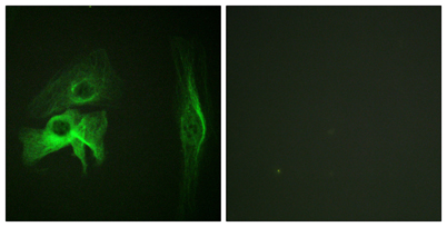
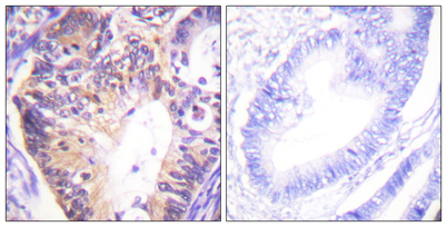
| Dilution | Western Blot: 1/500 - 1/2000. Immunohistochemistry: 1/100 - 1/300. Immunofluorescence: 1/200 - 1/1000. ELISA: 1/5000. Not yet tested in other applications. |
| Purification | The antibody was affinity-purified from rabbit antiserum by affinity-chromatography using epitope-specific immunogen. |
| Concentration | 1 mg/ml |
| Storage stability | -20°C/1 year |
| Molecular Weight (Da) | |
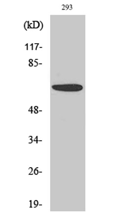
| Observed band (KD) | 60kD |
| Background | This gene encodes a family member of serine/threonine p21-activating kinases, known as PAK proteins. These proteins are critical effectors that link RhoGTPases to cytoskeleton reorganization and nuclear signaling, and they serve as targets for the small GTP binding proteins Cdc42 and Rac. This specific family member regulates cell motility and morphology. Alternatively spliced transcript variants encoding different isoforms have been found for this gene. [provided by RefSeq, Apr 2010], |
| Function | catalytic activity:ATP + a protein = ADP + a phosphoprotein.,cofactor:Magnesium.,enzyme regulation:Activated by binding small G proteins. Binding of GTP-bound CDC42 or RAC1 to the autoregulatory region releases monomers from the autoinhibited dimer, enables phosphorylation of Thr-423 and allows the kinase domain to adopt an active structure. Also activated by binding to GTP-bound CDC42, independent of the phosphorylation state of Thr-423. Phosphorylation of Thr-84 by OXSR1 inhibits this activation.,function:The activated kinase acts on a variety of targets. Likely to be the GTPase effector that links the Rho-related GTPases to the JNK MAP kinase pathway. Activated by CDC42 and RAC1. Involved in dissolution of stress fibers and reorganization of focal complexes. Involved in regulation of microtubule biogenesis through phosphorylation of TBCB. Activity is inhibited in cells undergoing apop |
| Subcellular location | Cytoplasm . Cell junction, focal adhesion . Cell projection, lamellipodium . Cell membrane . Cell projection, ruffle membrane . Cell projection, invadopodium . Nucleus, nucleoplasm . Chromosome . Cytoplasm, cytoskeleton, microtubule organizing center, centrosome . Colocalizes with RUFY3, F-actin and other core migration components in invadopodia at the cell periphery (PubMed:25766321). Recruited to the cell membrane by interaction with CDC42 and RAC1. Recruited to focal adhesions upon activation. Colocalized with CIB1 within membrane ruffles during cell spreading upon readhesion to fibronectin. Upon DNA damage, translocates to the nucleoplasm when phosphorylated at Thr-212 where is co-recruited with MORC2 on damaged chromatin (PubMed:23260667). Localization to the centrosome does not depen |
| Expression | Overexpressed in gastric cancer cells and tissues (at protein level) (PubMed:25766321). |

Use collapsible tabs for more detailed information that will help customers make a purchasing decision.
Ex: Shipping and return policies, size guides, and other common questions.