 Français
 English
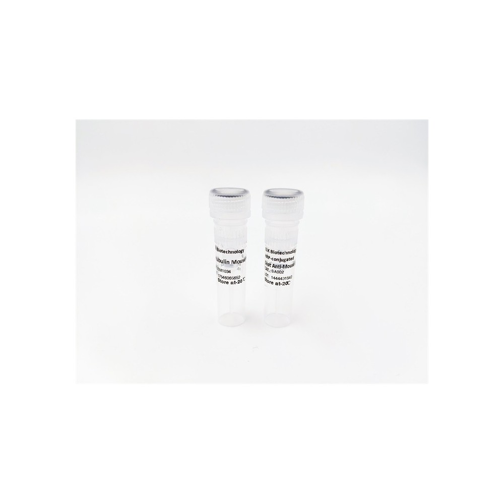

| ELK.No | ES3212 |
| Product name | PIPK I γ rabbit pAb |
| Reactivity | Human;Rat;Mouse; |
| Applications | WB;ELISA |
| Other name | PIP5K1C; KIAA0589; Phosphatidylinositol 4-phosphate 5-kinase type-1 gamma; PIP5K1-gamma; PtdIns(4)P-5-kinase 1 gamma; Phosphatidylinositol 4-phosphate 5-kinase type I gamma; PIP5KIgamma |
| Size | 50μL |
| Unit price ($) | 148 |
| Human gene ID | 23396 |
| Human Swiss-Prot | O60331 |
| Source | Rabbit |
| Isotype | IgG |
| Target | PIPK I γ |
| Fields | >>Inositol phosphate metabolism;>>Metabolic pathways;>>Phosphatidylinositol signaling system;>>Phospholipase D signaling pathway;>>Endocytosis;>>Focal adhesion;>>Fc gamma R-mediated phagocytosis;>>Regulation of actin cytoskeleton;>>Yersinia infection;>>Choline metabolism in cancer |
| Gene name | PIP5K1C |
| Protein name | Phosphatidylinositol 4-phosphate 5-kinase type-1 gamma |
| Human gene link | View Human Gene |
| Human Swiss link | View Human Swiss-Prot |
| Mouse gene ID | |
| Mouse gene link | |
| Mouse Swiss-Prot | O70161 |
| Mouse Swiss link | View Mouse Swiss-Prot |
| Rat gene ID | |
| Rat gene link | |
| Rat Swiss-Prot | |
| Rat Swiss link | |
| Immunogen | The antiserum was produced against synthesized peptide derived from human PIP5K1C. AA range:305-354 |
| Specificity | PIPK I γ Polyclonal Antibody detects endogenous levels of PIPK I γ protein. |
| Formulation | Liquid in PBS containing 50% glycerol, 0.5% BSA and 0.02% sodium azide. |
| Clonality | Polyclonal |
| Dilution | Western Blot: 1/500 - 1/2000. ELISA: 1/20000. Not yet tested in other applications. |
| Purification | The antibody was affinity-purified from rabbit antiserum by affinity-chromatography using epitope-specific immunogen. |
| Concentration | 1 mg/ml |
| Storage stability | -20°C/1 year |
| Molecular Weight (Da) | |
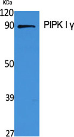
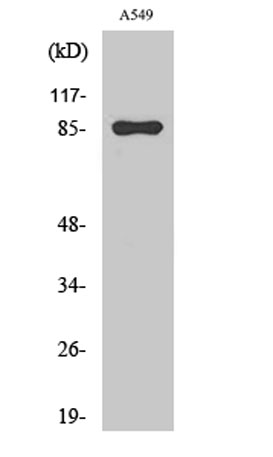
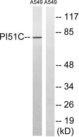
| Observed band (KD) | 80kD |
| Background | phosphatidylinositol-4-phosphate 5-kinase type 1 gamma(PIP5K1C) Homo sapiens This locus encodes a type I phosphatidylinositol 4-phosphate 5-kinase. The encoded protein catalyzes phosphorylation of phosphatidylinositol 4-phosphate, producing phosphatidylinositol 4,5-bisphosphate. This enzyme is found at synapses and has been found to play roles in endocytosis and cell migration. Mutations at this locus have been associated with lethal congenital contractural syndrome. Alternatively spliced transcript variants encoding different isoforms have been described.[provided by RefSeq, Sep 2010], |
| Function | catalytic activity:ATP + 1-phosphatidyl-1D-myo-inositol 4-phosphate = ADP + 1-phosphatidyl-1D-myo-inositol 4,5-bisphosphate.,disease:Defects in PIP5K1C are the cause of lethal congenital contractural syndrome type 3 (LCCS3) [MIM:611369]; also known as multiple contractural syndrome Israeli Bedouin type B. LCCS is an autosomal recessive disorder characterized by early fetal hydrops and akinesia, the Pena-Shokeir phenotype, specific neuropathology with degeneration of anterior horn neurons and extreme skeletal muscle atrophy. LCCS3 patients present at birth with severe multiple joint contractures with severe muscle wasting and atrophy, mainly in the legs. LCCS3 can be distinguished from the original LCCS by the absence of hydrops, fractures, and multiple pterygia.,enzyme regulation:Activated by interaction with TLN2.,function:Plays a role in membrane ruffling and assembly of clathrin-coate |
| Subcellular location | Cell membrane; Peripheral membrane protein; Cytoplasmic side . Endomembrane system . Cytoplasm . Cell junction, focal adhesion . Cell junction, adherens junction . Cell projection, ruffle membrane . Cell projection, phagocytic cup . Cell projection, uropodium . Detected in plasma membrane invaginations. Isoform 3 is detected in intracellular vesicle-like structures.; [Isoform 2]: Cytoplasm. Nucleus. |
| Expression | [Isoform 1]: Isoform 1 is strongly expressed in brain and also detected in heart and lung. ; [Isoform 2]: Isoform 2 is strongly expressed in pancreas and liver and in lesser quantities in brain, heart, lung and kidney. ; [Isoform 3]: Isoform 3 is detected in large amounts in heart and large intestine, is also present in lung, pancreas and thyroid, and to a lesser extent in brain, stomach and kidney. |

Use collapsible tabs for more detailed information that will help customers make a purchasing decision.
Ex: Shipping and return policies, size guides, and other common questions.