 Français
 English

| ELK.No | ES3338 |
| Product name | Renin Receptor rabbit pAb |
| Reactivity | Human;Mouse;Rat |
| Applications | WB;IHC;IF;ELISA |
| Other name | ATP6AP2; ATP6IP2; CAPER; ELDF10; HT028; MSTP009; PSEC0072; Renin receptor; ATPase H(+)-transporting lysosomal accessory protein 2; ATPase H(+)-transporting lysosomal-interacting protein 2; ER-localized type I transmembrane adaptor; Embryoni |
| Size | 100μL |
| Unit price ($) | 248 |
| Human gene ID | 10159 |
| Human Swiss-Prot | O75787 |
| Source | Rabbit |
| Isotype | IgG |
| Target | Renin Receptor |
| Fields | >>Renin-angiotensin system |
| Gene name | ATP6AP2 |
| Protein name | Renin receptor |
| Human gene link | View Human Gene |
| Human Swiss link | View Human Swiss-Prot |
| Mouse gene ID | 70495 |
| Mouse gene link | View Mouse Gene |
| Mouse Swiss-Prot | Q9CYN9 |
| Mouse Swiss link | View Mouse Swiss-Prot |
| Rat gene ID | 302526 |
| Rat gene link | View Rat Gene |
| Rat Swiss-Prot | Q6AXS4 |
| Rat Swiss link | View Rat Swiss-Prot |
| Immunogen | The antiserum was produced against synthesized peptide derived from human Caper. AA range:171-220 |
| Specificity | Renin Receptor Polyclonal Antibody detects endogenous levels of Renin Receptor protein. |
| Formulation | Liquid in PBS containing 50% glycerol, 0.5% BSA and 0.02% sodium azide. |
| Clonality | Polyclonal |
| Dilution | Western Blot: 1/500 - 1/2000. Immunohistochemistry: 1/100 - 1/300. Immunofluorescence: 1/200 - 1/1000. ELISA: 1/20000. Not yet tested in other applications. |
| Purification | The antibody was affinity-purified from rabbit antiserum by affinity-chromatography using epitope-specific immunogen. |
| Concentration | 1 mg/ml |
| Storage stability | -20°C/1 year |
| Molecular Weight (Da) | |
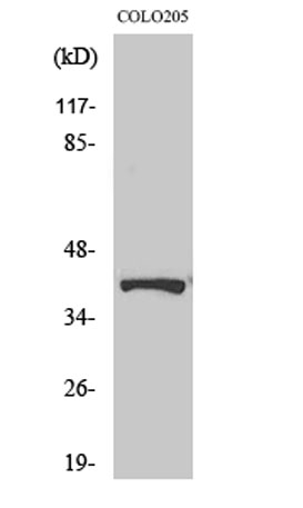
| Observed band (KD) | 39kD |
| Background | This gene encodes a protein that is associated with adenosine triphosphatases (ATPases). Proton-translocating ATPases have fundamental roles in energy conservation, secondary active transport, acidification of intracellular compartments, and cellular pH homeostasis. There are three classes of ATPases- F, P, and V. The vacuolar (V-type) ATPases have a transmembrane proton-conducting sector and an extramembrane catalytic sector. The encoded protein has been found associated with the transmembrane sector of the V-type ATPases. [provided by RefSeq, Jul 2008], |
| Function | disease:Defects in ATP6AP2 are a cause of mental retardation X-linked with epilepsy (MRXE) [MIM:300423]. MRXE is a syndromic mental retardation. Patients manifest mild to moderate mental retardation associated with epilepsy, delays in motor milestones and speech acquisition in infancy.,function:Functions as a renin and prorenin cellular receptor. May mediate renin-dependent cellular responses by activating ERK1 and ERK2. By increasing the catalytic efficiency of renin in AGT/angiotensinogen conversion to angiotensin I, it may also play a role in the renin-angiotensin system (RAS).,PTM:Phosphorylated.,subunit:Interacts with renin and the vacuolar proton-ATPase.,tissue specificity:Expressed in brain, heart, placenta, liver, kidney and pancreas. Barely detectable in lung and skeletal muscles. In the kidney cortex it is restricted to the mesangium of glomeruli. In the coronary and kidney art |
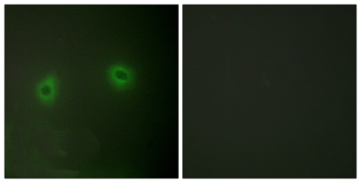
| Subcellular location | Endoplasmic reticulum membrane ; Single-pass type I membrane protein . Lysosome membrane ; Single-pass type I membrane protein . Cytoplasmic vesicle, autophagosome membrane ; Single-pass type I membrane protein . Cell projection, dendritic spine membrane ; Single-pass type I membrane protein . Cell projection, axon . Endosome membrane ; Single-pass type I membrane protein . Cytoplasmic vesicle, clathrin-coated vesicle membrane ; Single-pass type I membrane protein . Cytoplasmic vesicle, secretory vesicle, synaptic vesicle membrane ; Single-pass type I membrane protein . |
| Expression | Expressed in brain, heart, placenta, liver, kidney and pancreas. Barely detectable in lung and skeletal muscles. In the kidney cortex it is restricted to the mesangium of glomeruli. In the coronary and kidney artery it is expressed in the subendothelium, associated to smooth muscles where it colocalizes with REN. Expressed in vascular structures and by syncytiotrophoblast cells in the mature fetal placenta. |

Use collapsible tabs for more detailed information that will help customers make a purchasing decision.
Ex: Shipping and return policies, size guides, and other common questions.