 Français
 English

| ELK.No | ES3594 |
| Product name | TGFβ RI rabbit pAb |
| Reactivity | Human;Mouse;Rat |
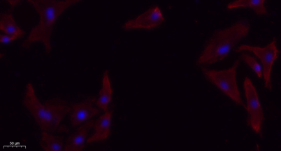
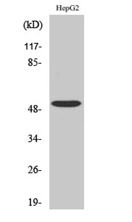
| Applications | WB;IF;ELISA |
| Other name | TGFBR1; ALK5; SKR4; TGF-beta receptor type-1; TGFR-1; Activin A receptor type II-like protein kinase of 53kD; Activin receptor-like kinase 5; ALK-5; ALK5; Serine/threonine-protein kinase receptor R4; SKR4; TGF-beta type I receptor; Transfor |
| Size | 50μL |
| Unit price ($) | 148 |
| Human gene ID | 7046 |
| Human Swiss-Prot | P36897 |
| Source | Rabbit |
| Isotype | IgG |
| Target | TGF β Receptor I |
| Fields | >>MAPK signaling pathway;>>Cytokine-cytokine receptor interaction;>>FoxO signaling pathway;>>Endocytosis;>>Cellular senescence;>>TGF-beta signaling pathway;>>Apelin signaling pathway;>>Osteoclast differentiation;>>Hippo signaling pathway;>>Adherens junction;>>Th17 cell differentiation;>>Relaxin signaling pathway;>>AGE-RAGE signaling pathway in diabetic complications;>>Chagas disease;>>Hepatitis B;>>Human T-cell leukemia virus 1 infection;>>Pathways in cancer;>>Colorectal cancer;>>Pancreatic cancer;>>Chronic myeloid leukemia;>>Hepatocellular carcinoma;>>Gastric cancer;>>Diabetic cardiomyopathy |
| Gene name | TGFBR1 |
| Protein name | TGF-beta receptor type-1 |
| Human gene link | View Human Gene |
| Human Swiss link | View Human Swiss-Prot |
| Mouse gene ID | 21812 |
| Mouse gene link | View Mouse Gene |
| Mouse Swiss-Prot | Q64729 |
| Mouse Swiss link | View Mouse Swiss-Prot |
| Rat gene ID | |
| Rat gene link | |
| Rat Swiss-Prot | P80204 |
| Rat Swiss link | View Rat Swiss-Prot |
| Immunogen | The antiserum was produced against synthesized peptide derived from human TGF beta Receptor I. AA range:131-180 |
| Specificity | TGFβ RI Polyclonal Antibody detects endogenous levels of TGFβ RI protein. |
| Formulation | Liquid in PBS containing 50% glycerol, 0.5% BSA and 0.02% sodium azide. |
| Clonality | Polyclonal |
| Dilution | Western Blot: 1/500 - 1/2000. IHC-p: 1:100-300 ELISA: 1/20000. IF 1:100-300 Not yet tested in other applications. |
| Purification | The antibody was affinity-purified from rabbit antiserum by affinity-chromatography using epitope-specific immunogen. |
| Concentration | 1 mg/ml |
| Storage stability | -20°C/1 year |
| Molecular Weight (Da) | |
| Observed band (KD) | 56kD |
| Background | The protein encoded by this gene forms a heteromeric complex with type II TGF-beta receptors when bound to TGF-beta, transducing the TGF-beta signal from the cell surface to the cytoplasm. The encoded protein is a serine/threonine protein kinase. Mutations in this gene have been associated with Loeys-Dietz aortic aneurysm syndrome (LDAS). Multiple transcript variants encoding different isoforms have been found for this gene. [provided by RefSeq, Aug 2008], |
| Function | catalytic activity:ATP + [receptor-protein] = ADP + [receptor-protein] phosphate.,cofactor:Magnesium or manganese.,disease:Defects in TGFBR1 are the cause of aortic aneurysm familial thoracic type 5 (AAT5) [MIM:608967]. Aneurysms and dissections of the aorta usually result from degenerative changes in the aortic wall. Thoracic aortic aneurysms and dissections are primarily associated with a characteristic histologic appearance known as 'medial necrosis' in which there is degeneration and fragmentation of elastic fibers, loss of smooth muscle cells, and an accumulation of basophilic ground substance.,disease:Defects in TGFBR1 are the cause of Loeys-Dietz syndrome type 1A (LDS1A) [MIM:609192]; also known as Furlong syndrome or Loeys-Dietz aortic aneurysm syndrome (LDAS). LDS1 is an aortic aneurysm syndrome with widespread systemic involvement. The disorder is characterized by arterial tort |
| Subcellular location | Cell membrane ; Single-pass type I membrane protein . Cell junction, tight junction . Cell surface . Membrane raft . |
| Expression | Found in all tissues examined, most abundant in placenta and least abundant in brain and heart. Expressed in a variety of cancer cell lines (PubMed:25893292). |


Use collapsible tabs for more detailed information that will help customers make a purchasing decision.
Ex: Shipping and return policies, size guides, and other common questions.