 Français
 English

| ELK.No | ES3622 |
| Product name | TNF-R2 rabbit pAb |
| Reactivity | Human;Mouse;Rat |
| Applications | WB;IHC;IF;ELISA |
| Other name | TNFRSF1B; TNFBR; TNFR2; Tumor necrosis factor receptor superfamily member 1B; Tumor necrosis factor receptor 2; TNF-R2; Tumor necrosis factor receptor type II; TNF-RII; TNFR-II; p75; p80 TNF-alpha receptor; CD antigen CD120b; Etanercept |
| Size | 100μL |
| Unit price ($) | 248 |
| Human gene ID | 7133 |
| Human Swiss-Prot | P20333 |
| Source | Rabbit |
| Isotype | IgG |
| Target | TNF-R2 |
| Fields | >>Cytokine-cytokine receptor interaction;>>Viral protein interaction with cytokine and cytokine receptor;>>TNF signaling pathway;>>Adipocytokine signaling pathway;>>Amyotrophic lateral sclerosis;>>Pathways of neurodegeneration - multiple diseases;>>Human immunodeficiency virus 1 infection |
| Gene name | TNFRSF1B |
| Protein name | Tumor necrosis factor receptor superfamily member 1B |
| Human gene link | View Human Gene |
| Human Swiss link | View Human Swiss-Prot |
| Mouse gene ID | 21938 |
| Mouse gene link | View Mouse Gene |
| Mouse Swiss-Prot | P25119 |
| Mouse Swiss link | View Mouse Swiss-Prot |
| Rat gene ID | 156767 |
| Rat gene link | View Rat Gene |
| Rat Swiss-Prot | Q80WY6 |
| Rat Swiss link | View Rat Swiss-Prot |
| Immunogen | The antiserum was produced against synthesized peptide derived from human TNF Receptor II. AA range:376-425 |
| Specificity | TNF-R2 Polyclonal Antibody detects endogenous levels of TNF-R2 protein. |
| Formulation | Liquid in PBS containing 50% glycerol, 0.5% BSA and 0.02% sodium azide. |
| Clonality | Polyclonal |
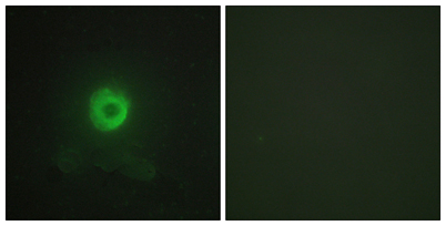
| Dilution | Western Blot: 1/500 - 1/2000. Immunohistochemistry: 1/100 - 1/300. Immunofluorescence: 1/200 - 1/1000. ELISA: 1/20000. Not yet tested in other applications. |
| Purification | The antibody was affinity-purified from rabbit antiserum by affinity-chromatography using epitope-specific immunogen. |
| Concentration | 1 mg/ml |
| Storage stability | -20°C/1 year |
| Molecular Weight (Da) | |
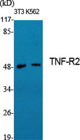
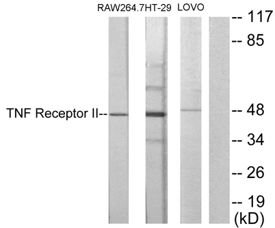
| Observed band (KD) | 48kD |
| Background | The protein encoded by this gene is a member of the TNF-receptor superfamily. This protein and TNF-receptor 1 form a heterocomplex that mediates the recruitment of two anti-apoptotic proteins, c-IAP1 and c-IAP2, which possess E3 ubiquitin ligase activity. The function of IAPs in TNF-receptor signalling is unknown, however, c-IAP1 is thought to potentiate TNF-induced apoptosis by the ubiquitination and degradation of TNF-receptor-associated factor 2, which mediates anti-apoptotic signals. Knockout studies in mice also suggest a role of this protein in protecting neurons from apoptosis by stimulating antioxidative pathways. [provided by RefSeq, Jul 2008], |
| Function | function:Receptor with high affinity for TNFSF2/TNF-alpha and approximately 5-fold lower affinity for homotrimeric TNFSF1/lymphotoxin-alpha. The TRAF1/TRAF2 complex recruits the apoptotic suppressors BIRC2 and BIRC3 to TNFRSF1B/TNFR2. This receptor mediates most of the metabolic effects of TNF-alpha. Isoform 2 blocks TNF-alpha-induced apoptosis, which suggests that it regulates TNF-alpha function by antagonizing its biological activity.,online information:Clinical information on Enbrel,pharmaceutical:Available under the name Enbrel (Immunex and Wyeth-Ayerst). Used to treat moderate to severe rheumatoid arthritis (RA). Enbrel consist of the extracellular ligand-binding portion of TNFRSF1B linked to an immunoglobulin Fc chain. It binds to TNF-alpha and blocks its interactions with receptors.,PTM:A soluble form (tumor necrosis factor binding protein 2) is produced from the membrane form by |
| Subcellular location | [Isoform 1]: Cell membrane; Single-pass type I membrane protein.; [Isoform 2]: Secreted.; [Tumor necrosis factor-binding protein 2]: Secreted. |
| Expression | Muscle,PNS,Urine, |

Use collapsible tabs for more detailed information that will help customers make a purchasing decision.
Ex: Shipping and return policies, size guides, and other common questions.