 Français
 English
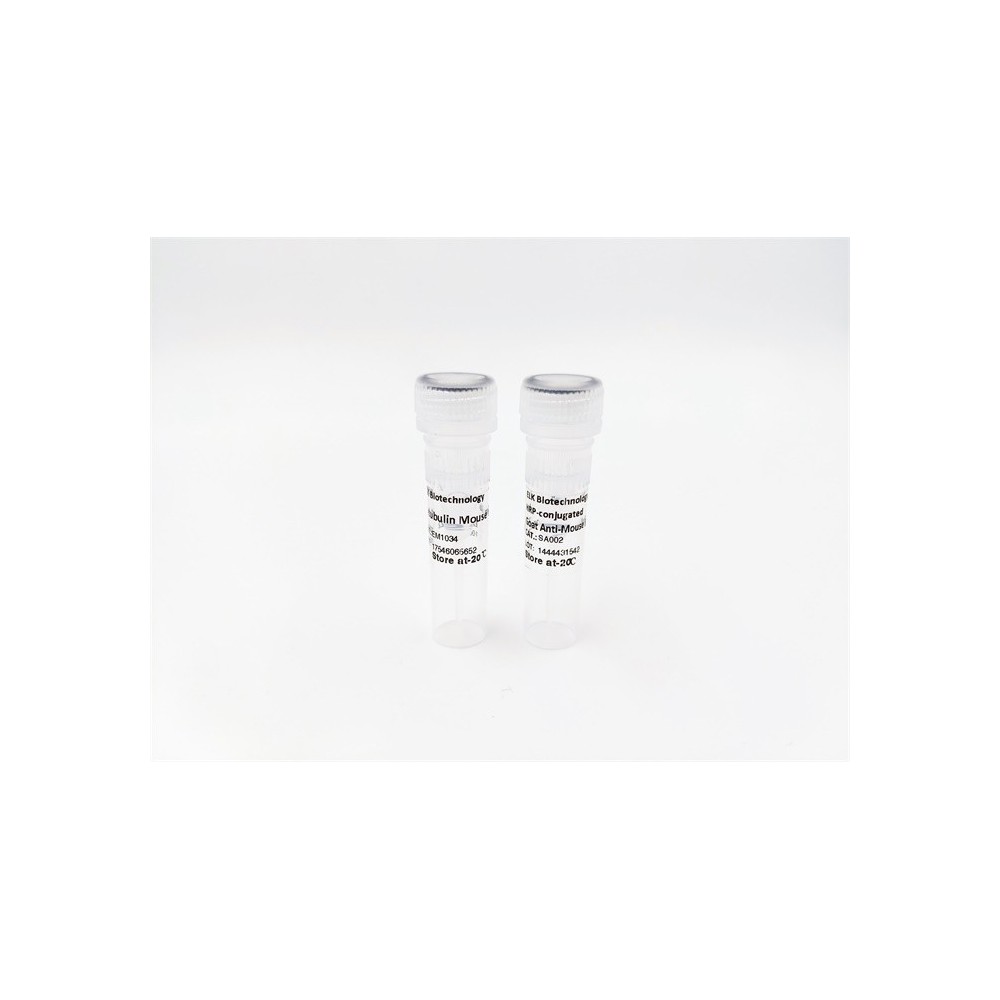

| ELK.No | ES3702 |
| Product name | WAVE1 rabbit pAb |
| Reactivity | Human;Mouse;Rat |
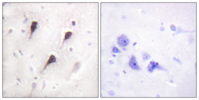
| Applications | WB;IHC;IF;ELISA |
| Other name | WASF1; KIAA0269; SCAR1; WAVE1; Wiskott-Aldrich syndrome protein family member 1; WASP family protein member 1; Protein WAVE-1; Verprolin homology domain-containing protein 1 |
| Size | 50μL |
| Unit price ($) | 148 |
| Human gene ID | 8936 |
| Human Swiss-Prot | Q92558 |
| Source | Rabbit |
| Isotype | IgG |
| Target | WAVE1 |
| Fields | >>Adherens junction;>>Fc gamma R-mediated phagocytosis;>>Regulation of actin cytoskeleton;>>Bacterial invasion of epithelial cells;>>Pathogenic Escherichia coli infection;>>Shigellosis;>>Choline metabolism in cancer |
| Gene name | WASF1 |
| Protein name | Wiskott-Aldrich syndrome protein family member 1 |
| Human gene link | View Human Gene |
| Human Swiss link | View Human Swiss-Prot |
| Mouse gene ID | 83767 |
| Mouse gene link | View Mouse Gene |
| Mouse Swiss-Prot | Q8R5H6 |
| Mouse Swiss link | View Mouse Swiss-Prot |
| Rat gene ID | 294568 |
| Rat gene link | View Rat Gene |
| Rat Swiss-Prot | Q5BJU7 |
| Rat Swiss link | View Rat Swiss-Prot |
| Immunogen | The antiserum was produced against synthesized peptide derived from human WAVE1. AA range:91-140 |
| Specificity | WAVE1 Polyclonal Antibody detects endogenous levels of WAVE1 protein. |
| Formulation | Liquid in PBS containing 50% glycerol, 0.5% BSA and 0.02% sodium azide. |
| Clonality | Polyclonal |
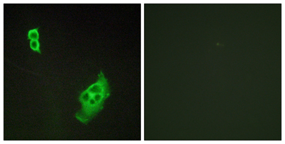
| Dilution | Western Blot: 1/500 - 1/2000. Immunohistochemistry: 1/100 - 1/300. Immunofluorescence: 1/200 - 1/1000. ELISA: 1/40000. Not yet tested in other applications. |
| Purification | The antibody was affinity-purified from rabbit antiserum by affinity-chromatography using epitope-specific immunogen. |
| Concentration | 1 mg/ml |
| Storage stability | -20°C/1 year |
| Molecular Weight (Da) | |
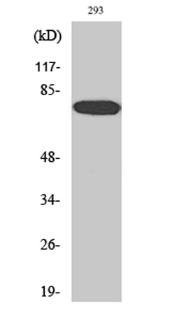
| Observed band (KD) | 70kD |
| Background | The protein encoded by this gene, a member of the Wiskott-Aldrich syndrome protein (WASP)-family, plays a critical role downstream of Rac, a Rho-family small GTPase, in regulating the actin cytoskeleton required for membrane ruffling. It has been shown to associate with an actin nucleation core Arp2/3 complex while enhancing actin polymerization in vitro. Wiskott-Aldrich syndrome is a disease of the immune system, likely due to defects in regulation of actin cytoskeleton. Multiple alternatively spliced transcript variants encoding the same protein have been found for this gene. [provided by RefSeq, Jul 2008], |
| Function | domain:Binds the Arp2/3 complex through the C-terminal region and actin through verprolin homology (VPH) domain.,function:Downstream effector molecules involved in the transmission of signals from tyrosine kinase receptors and small GTPases to the actin cytoskeleton.,similarity:Belongs to the SCAR/WAVE family.,similarity:Contains 1 WH2 domain.,subcellular location:Dot-like pattern in the cytoplasm. Concentrated in Rac-regulated membrane-ruffling areas.,subunit:Component of the WAVE1 complex composed of ABI2, CYFIP2, C3orf10/HSPC300, NCKAP1 and WASF1/WAVE1. CYFIP2 binds to activated RAC1 which causes the complex to dissociate, releasing activated WASF1. The complex can also be activated by NCK1 (By similarity). Binds actin and the Arp2/3 complex. Interacts with BAIAP2.,tissue specificity:Highly expressed in brain. Lowly expressed in testis, ovary, colon, kidney, pancreas, thymus, small in |
| Subcellular location | Cytoplasm, cytoskeleton . Cell junction, synapse . Cell junction, focal adhesion . Dot-like pattern in the cytoplasm. Concentrated in Rac-regulated membrane-ruffling areas (PubMed:9889097). Partial translocation to focal adhesion sites might be mediated by interaction with SORBS2 (PubMed:18559503). In neurons, colocalizes with activated NTRK2 after BDNF addition in endocytic sites through the association with TMEM108 (By similarity). . |
| Expression | Highly expressed in brain. Lowly expressed in testis, ovary, colon, kidney, pancreas, thymus, small intestine and peripheral blood. |

Use collapsible tabs for more detailed information that will help customers make a purchasing decision.
Ex: Shipping and return policies, size guides, and other common questions.