 Français
 English
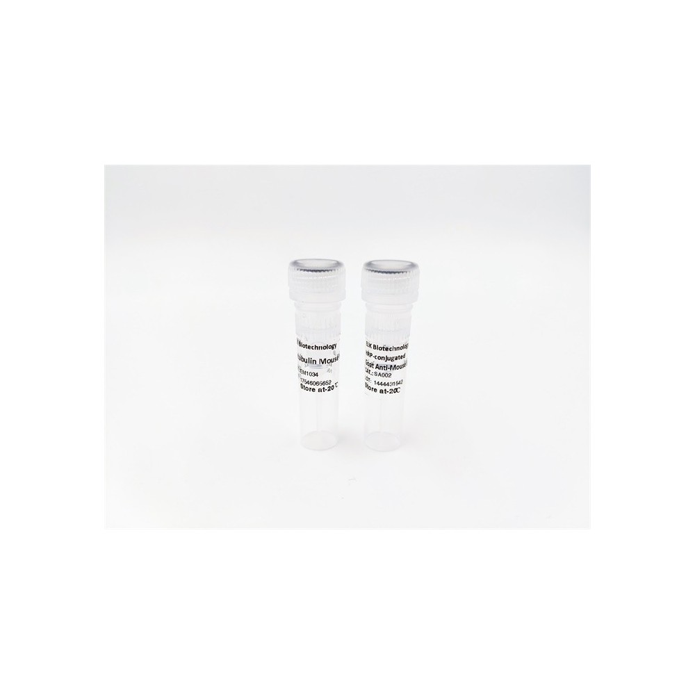

| ELK.No | ES3802 |
| Product name | Myosin VI rabbit pAb |
| Reactivity | Human;Mouse;Rat |
| Applications | WB;IHC;IF;ELISA |
| Other name | MYO6; KIAA0389; Unconventional myosin-VI; Unconventional myosin-6 |
| Size | 100μL |
| Unit price ($) | 248 |
| Human gene ID | 4646 |
| Human Swiss-Prot | Q9UM54 |
| Source | Rabbit |
| Isotype | IgG |
| Target | Myosin VI |
| Fields | >>Pathogenic Escherichia coli infection;>>Salmonella infection |
| Gene name | MYO6 |
| Protein name | Unconventional myosin-VI |
| Human gene link | View Human Gene |
| Human Swiss link | View Human Swiss-Prot |
| Mouse gene ID | |
| Mouse gene link | |
| Mouse Swiss-Prot | Q64331 |
| Mouse Swiss link | View Mouse Swiss-Prot |
| Rat gene ID | |
| Rat gene link | |
| Rat Swiss-Prot | |
| Rat Swiss link | |
| Immunogen | Synthesized peptide derived from Myosin VI . at AA range: 40-120 |
| Specificity | Myosin VI Polyclonal Antibody detects endogenous levels of Myosin VI protein. |
| Formulation | Liquid in PBS containing 50% glycerol, 0.5% BSA and 0.02% sodium azide. |
| Clonality | Polyclonal |
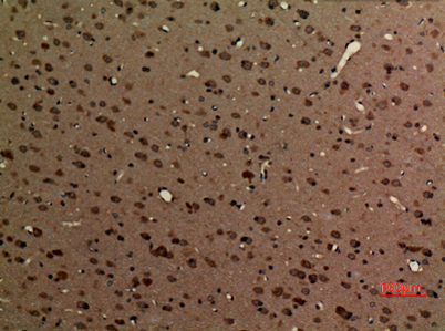
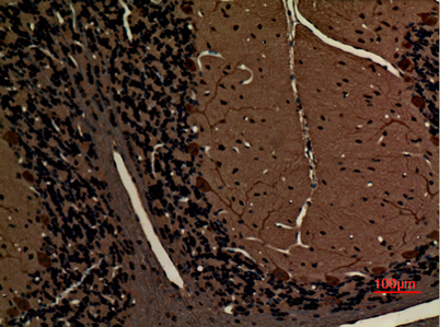
| Dilution | Western Blot: 1/500 - 1/2000. IHC-p: 1:100-300 ELISA: 1/5000. Not yet tested in other applications. |
| Purification | The antibody was affinity-purified from rabbit antiserum by affinity-chromatography using epitope-specific immunogen. |
| Concentration | 1 mg/ml |
| Storage stability | -20°C/1 year |
| Molecular Weight (Da) | |
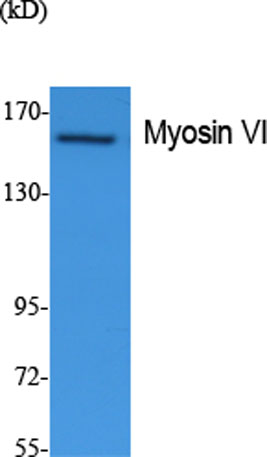
| Observed band (KD) | 149kD |
| Background | myosin VI(MYO6) Homo sapiens This gene encodes a reverse-direction motor protein that moves toward the minus end of actin filaments and plays a role in intracellular vesicle and organelle transport. The protein consists of a motor domain containing an ATP- and an actin-binding site and a globular tail which interacts with other proteins. This protein maintains the structural integrity of inner ear hair cells and mutations in this gene cause non-syndromic autosomal dominant and recessive hearing loss. Alternative splicing results in multiple transcript variants encoding distinct isoforms. [provided by RefSeq, Jul 2014], |
| Function | disease:Defects in MYO6 are the cause of non-syndromic sensorineural deafness autosomal dominant type 22 (DFNA22) [MIM:606346]. DFNA22 is a form of sensorineural hearing loss. Sensorineural deafness results from damage to the neural receptors of the inner ear, the nerve pathways to the brain, or the area of the brain that receives sound information. DFNA22 is progressive and postlingual, with onset during childhood. By the age of approximately 50 years, affected individuals invariably have profound sensorineural deafness.,disease:Defects in MYO6 are the cause of non-syndromic sensorineural deafness autosomal recessive type 37 (DFNB37) [MIM:607821].,disease:Defects in MYO6 are the cause of sensorineural deafness with hypertrophic cardiomyopathy (DFNHCM) [MIM:606346].,domain:Divided into three regions: a N-terminal motor (head) domain, followed by a neck domain consisting of a calmodulin-b |
| Subcellular location | Golgi apparatus, trans-Golgi network membrane ; Peripheral membrane protein . Golgi apparatus . Nucleus . Cytoplasm, perinuclear region . Membrane, clathrin-coated pit . Cytoplasmic vesicle, clathrin-coated vesicle . Cell projection, filopodium . Cell projection, ruffle membrane . Cell projection, microvillus . Cytoplasm, cytosol . Also present in endocyctic vesicles (PubMed:16507995). Translocates from membrane ruffles, endocytic vesicles and cytoplasm to Golgi apparatus, perinuclear membrane and nucleus through induction by p53 and p53-induced DNA damage (PubMed:16507995). Recruited into membrane ruffles from cell surface by EGF-stimulation (PubMed:9852149). Colocalizes with DAB2 in clathrin-coated pits/vesicles (PubMed:11967127). Colocalizes with OPTN at the Golgi complex and in vesicul |
| Expression | Expressed in most tissues examined including heart, brain, placenta, pancreas, spleen, thymus, prostate, testis, ovary, small intestine and colon. Highest levels in brain, pancreas, testis and small intestine. Also expressed in fetal brain and cochlea. Isoform 1 and isoform 2, containing the small insert, and isoform 4, containing neither insert, are expressed in unpolarized epithelial cells. |

Use collapsible tabs for more detailed information that will help customers make a purchasing decision.
Ex: Shipping and return policies, size guides, and other common questions.