 Français
 English

| ELK.No | ES4029 |
| Product name | PDGF-D rabbit pAb |
| Reactivity | Human;Rat;Mouse; |
| Applications | WB;IHC;IF;ELISA |
| Other name | PDGFD; IEGF; SCDGFB; MSTP036; Platelet-derived growth factor D; PDGF-D; Iris-expressed growth factor; Spinal cord-derived growth factor B; SCDGF-B |
| Size | 50μL |
| Unit price ($) | 148 |
| Human gene ID | 80310 |
| Human Swiss-Prot | Q9GZP0 |
| Source | Rabbit |
| Isotype | IgG |
| Target | PDGF-D |
| Fields | >>EGFR tyrosine kinase inhibitor resistance;>>MAPK signaling pathway;>>Ras signaling pathway;>>Rap1 signaling pathway;>>Calcium signaling pathway;>>Phospholipase D signaling pathway;>>PI3K-Akt signaling pathway;>>Focal adhesion;>>Gap junction;>>Regulation of actin cytoskeleton;>>Prostate cancer;>>Melanoma;>>Choline metabolism in cancer |
| Gene name | PDGFD |
| Protein name | Platelet-derived growth factor D |
| Human gene link | View Human Gene |
| Human Swiss link | View Human Swiss-Prot |
| Mouse gene ID | 71785 |
| Mouse gene link | View Mouse Gene |
| Mouse Swiss-Prot | Q925I7 |
| Mouse Swiss link | View Mouse Swiss-Prot |
| Rat gene ID | 66018 |
| Rat gene link | View Rat Gene |
| Rat Swiss-Prot | Q9EQT1 |
| Rat Swiss link | View Rat Swiss-Prot |
| Immunogen | The antiserum was produced against synthesized peptide derived from the C-terminal region of human PDGFD. AA range:311-360 |
| Specificity | PDGF-D Polyclonal Antibody detects endogenous levels of PDGF-D protein. |
| Formulation | Liquid in PBS containing 50% glycerol, 0.5% BSA and 0.02% sodium azide. |
| Clonality | Polyclonal |
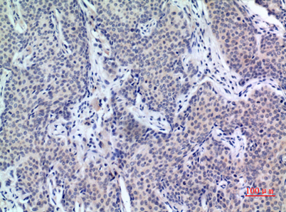
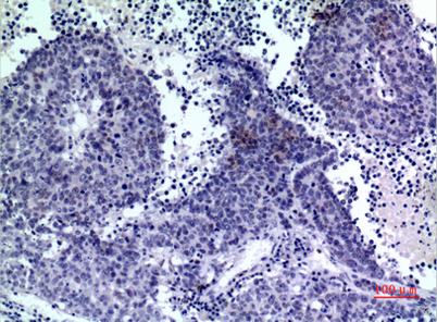
| Dilution | Western Blot: 1/500 - 1/2000. IHC-p: 1/100-1/300. ELISA: 1/20000. Not yet tested in other applications. |
| Purification | The antibody was affinity-purified from rabbit antiserum by affinity-chromatography using epitope-specific immunogen. |
| Concentration | 1 mg/ml |
| Storage stability | -20°C/1 year |
| Molecular Weight (Da) | |
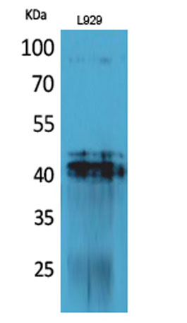
| Observed band (KD) | 42kD |
| Background | platelet derived growth factor D(PDGFD) Homo sapiens The protein encoded by this gene is a member of the platelet-derived growth factor family. The four members of this family are mitogenic factors for cells of mesenchymal origin and are characterized by a core motif of eight cysteines, seven of which are found in this factor. This gene product only forms homodimers and, therefore, does not dimerize with the other three family members. It differs from alpha and beta members of this family in having an unusual N-terminal domain, the CUB domain. Two splice variants have been identified for this gene. [provided by RefSeq, Jul 2008], |
| Function | developmental stage:Not detectable in the earliest stages of glomerulogenesis, and not detected in the metanephric blastema or surrounding cortical interstitial cells. In later stages of glomerulogenesis, localized to epithelial cells transitioning from the early developing nephrons of the comma- and S-shaped stages to the visceral epithelial cells of differentiated glomeruli. In the developing pelvis, expressed at the basement membrane of immature collecting ducts and by presumptive fibroblastic cells in the interstitium.,function:Potent mitogen for cells of mesenchymal origin. Binding of this growth factor to its affinity receptor elicits a variety of cellular responses. It is released by platelets upon wounding and plays an important role in stimulating adjacent cells to grow and thereby heals the wound. Activated by proteolytic cleavage and this active form acts as a specific ligand |
| Subcellular location | Secreted . Released by platelets upon wounding. |
| Expression | Expressed at high levels in the heart, pancreas, adrenal gland and ovary and at low levels in placenta, liver, kidney, prostate, testis, small intestine, spleen and colon. In the kidney, expressed by the visceral epithelial cells of the glomeruli. A widespread expression is also seen in the medial smooth muscle cells of arteries and arterioles, as well as in smooth muscle cells of vasa rectae in the medullary area. Expressed in the adventitial connective tissue surrounding the suprarenal artery. In chronic obstructive nephropathy, a persistent expression is seen in glomerular visceral epithelial cells and vascular smooth muscle cells, as well as de novo expression by periglomerular interstitial cells and by some neointimal cells of atherosclerotic vessels. Expression in normal prostate is |

Use collapsible tabs for more detailed information that will help customers make a purchasing decision.
Ex: Shipping and return policies, size guides, and other common questions.