 Français
 English

| ELK.No | ES4079 |
| Product name | Cytokeratin 19 rabbit pAb |
| Reactivity | Human;Rat;Mouse; |
| Applications | WB;IHC;IF;ELISA |
| Other name | KRT19; Keratin, type I cytoskeletal 19; Cytokeratin-19; CK-19; Keratin-19; K19 |
| Size | 100μL |
| Unit price ($) | 248 |
| Human gene ID | 3880 |
| Human Swiss-Prot | P08727 |
| Source | Rabbit |
| Isotype | IgG |
| Target | Cytokeratin 19 |
| Fields | >>Estrogen signaling pathway;>>Staphylococcus aureus infection |
| Gene name | KRT19 |
| Protein name | Keratin type I cytoskeletal 19 |
| Human gene link | View Human Gene |
| Human Swiss link | View Human Swiss-Prot |
| Mouse gene ID | |
| Mouse gene link | |
| Mouse Swiss-Prot | P19001 |
| Mouse Swiss link | View Mouse Swiss-Prot |
| Rat gene ID | |
| Rat gene link | |
| Rat Swiss-Prot | |
| Rat Swiss link | |
| Immunogen | The antiserum was produced against synthesized peptide derived from the C-terminal region of human KRT19. AA range:321-370 |
| Specificity | Cytokeratin 19 Polyclonal Antibody detects endogenous levels of Cytokeratin 19 protein. |
| Formulation | Liquid in PBS containing 50% glycerol, 0.5% BSA and 0.02% sodium azide. |
| Clonality | Polyclonal |
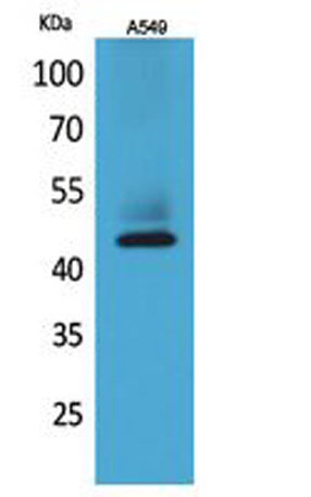
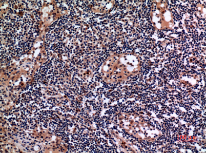
| Dilution | Western Blot: 1/500 - 1/2000. IHC-p: 1/100-1/300. ELISA: 1/20000. Not yet tested in other applications. |
| Purification | The antibody was affinity-purified from rabbit antiserum by affinity-chromatography using epitope-specific immunogen. |
| Concentration | 1 mg/ml |
| Storage stability | -20°C/1 year |
| Molecular Weight (Da) | |
| Observed band (KD) | 44kD |
| Background | The protein encoded by this gene is a member of the keratin family. The keratins are intermediate filament proteins responsible for the structural integrity of epithelial cells and are subdivided into cytokeratins and hair keratins. The type I cytokeratins consist of acidic proteins which are arranged in pairs of heterotypic keratin chains. Unlike its related family members, this smallest known acidic cytokeratin is not paired with a basic cytokeratin in epithelial cells. It is specifically expressed in the periderm, the transiently superficial layer that envelopes the developing epidermis. The type I cytokeratins are clustered in a region of chromosome 17q12-q21. [provided by RefSeq, Jul 2008], |
| Function | developmental stage:Present in hair follicles at all stages of development.,domain:This keratin differs from all other IF proteins in lacking the C-terminal tail domain.,function:Involved in the organization of myofibers. Together with KRT8, helps to link the contractile apparatus to dystrophin at the costameres of striated muscle.,miscellaneous:There are two types of cytoskeletal and microfibrillar keratin: I (acidic; 40-55 kDa) and II (neutral to basic; 56-70 kDa).,similarity:Belongs to the intermediate filament family.,subunit:Heterotetramer of two type I and two type II keratins. Interacts with PNN and the actin-binding domain of DMD. Interacts with HCV core protein.,tissue specificity:Expressed in a defined zone of basal keratinocytes in the deep outer root sheath of hair follicles. Also observed in sweat gland and mammary gland ductal and secretory cells, bile ducts, gastrointestin |
| Subcellular location | intermediate filament,plasma membrane,dystrophin-associated glycoprotein complex,Z disc,sarcolemma,costamere,extracellular exosome,cell periphery,terminal web, |
| Expression | Expressed in a defined zone of basal keratinocytes in the deep outer root sheath of hair follicles. Also observed in sweat gland and mammary gland ductal and secretory cells, bile ducts, gastrointestinal tract, bladder urothelium, oral epithelia, esophagus, ectocervical epithelium (at protein level). Expressed in epidermal basal cells, in nipple epidermis and a defined region of the hair follicle. Also seen in a subset of vascular wall cells in both the veins and artery of human umbilical cord, and in umbilical cord vascular smooth muscle. Observed in muscle fibers accumulating in the costameres of myoplasm at the sarcolemma in structures that contain dystrophin and spectrin. |

Use collapsible tabs for more detailed information that will help customers make a purchasing decision.
Ex: Shipping and return policies, size guides, and other common questions.