 Français
 English
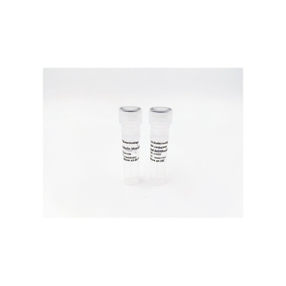

| ELK.No | ES4090 |
| Product name | Bag-1 rabbit pAb |
| Reactivity | Human;Rat;Mouse; |
| Applications | WB;ELISA |
| Other name | BAG1; HAP; BAG family molecular chaperone regulator 1; BAG-1; Bcl-2-associated athanogene 1 |
| Size | 50μL |
| Unit price ($) | 148 |
| Human gene ID | 573 |
| Human Swiss-Prot | Q99933 |
| Source | Rabbit |
| Isotype | IgG |
| Target | Bag-1 |
| Fields | >>Protein processing in endoplasmic reticulum |
| Gene name | BAG1 |
| Protein name | BAG family molecular chaperone regulator 1 |
| Human gene link | View Human Gene |
| Human Swiss link | View Human Swiss-Prot |
| Mouse gene ID | |
| Mouse gene link | |
| Mouse Swiss-Prot | Q60739 |
| Mouse Swiss link | View Mouse Swiss-Prot |
| Rat gene ID | |
| Rat gene link | |
| Rat Swiss-Prot | |
| Rat Swiss link | |
| Immunogen | The antiserum was produced against synthesized peptide derived from the Internal region of human BAG1. AA range:41-90 |
| Specificity | Bag-1 Polyclonal Antibody detects endogenous levels of Bag-1 protein. |
| Formulation | Liquid in PBS containing 50% glycerol, 0.5% BSA and 0.02% sodium azide. |
| Clonality | Polyclonal |
| Dilution | Western Blot: 1/500 - 1/2000. ELISA: 1/20000. Not yet tested in other applications. |
| Purification | The antibody was affinity-purified from rabbit antiserum by affinity-chromatography using epitope-specific immunogen. |
| Concentration | 1 mg/ml |
| Storage stability | -20°C/1 year |
| Molecular Weight (Da) | |
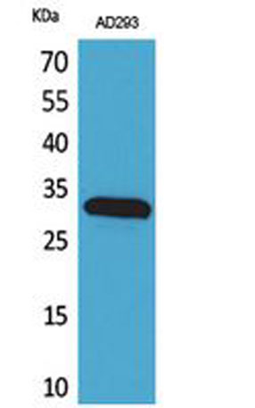
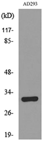
| Observed band (KD) | 35kD |
| Background | The oncogene BCL2 is a membrane protein that blocks a step in a pathway leading to apoptosis or programmed cell death. The protein encoded by this gene binds to BCL2 and is referred to as BCL2-associated athanogene. It enhances the anti-apoptotic effects of BCL2 and represents a link between growth factor receptors and anti-apoptotic mechanisms. Multiple protein isoforms are encoded by this mRNA through the use of a non-AUG (CUG) initiation codon, and three alternative downstream AUG initiation codons. A related pseudogene has been defined on chromosome X. [provided by RefSeq, Feb 2010], |
| Function | disease:May be linked to the cryptophthalmos syndrome (Fraser syndrome), an autosomal recessive disorder characterized by the failure of eyes fissures to form during embryogenesis, webbed fingers, and atresia of ear canals, anus, vagina, alimentary tract, or larynx. All these developmental processes require cell death.,function:Inhibits the chaperone activity of HSP70/HSC70 by promoting substrate release. Inhibits the pro-apoptotic function of PPP1R15A, and has anti-apoptotic activity. Markedly increases the anti-cell death function of BCL2 induced by various stimuli.,PTM:Ubiquitinated; mediated by SIAH1 or SIAH2 and leading to its subsequent proteasomal degradation.,similarity:Contains 1 BAG domain.,similarity:Contains 1 ubiquitin-like domain.,subcellular location:Isoform2 localizes to the cytoplasm and shuttles into the nucleus in response to heat shock.,subunit:Binds to the ATPase dom |
| Subcellular location | [Isoform 1]: Nucleus. Cytoplasm. Isoform 1 localizes predominantly to the nucleus.; [Isoform 2]: Cytoplasm. Nucleus. Isoform 2 localizes to the cytoplasm and shuttles into the nucleus in response to heat shock.; [Isoform 4]: Cytoplasm. Nucleus. Isoform 4 localizes predominantly to the cytoplasm. The cellular background in which it is expressed can influence whether it resides primarily in the cytoplasm or is also found in the nucleus. In the presence of BCL2, localizes to intracellular membranes (what appears to be the nuclear envelope and perinuclear membranes) as well as punctate cytosolic structures suggestive of mitochondria. |
| Expression | Isoform 4 is the most abundantly expressed isoform. It is ubiquitously expressed throughout most tissues, except the liver, colon, breast and uterine myometrium. Isoform 1 is expressed in the ovary and testis. Isoform 4 is expressed in several types of tumor cell lines, and at consistently high levels in leukemia and lymphoma cell lines. Isoform 1 is expressed in the prostate, breast and leukemia cell lines. Isoform 3 is the least abundant isoform in tumor cell lines (at protein level). |

Use collapsible tabs for more detailed information that will help customers make a purchasing decision.
Ex: Shipping and return policies, size guides, and other common questions.