 Français
 English
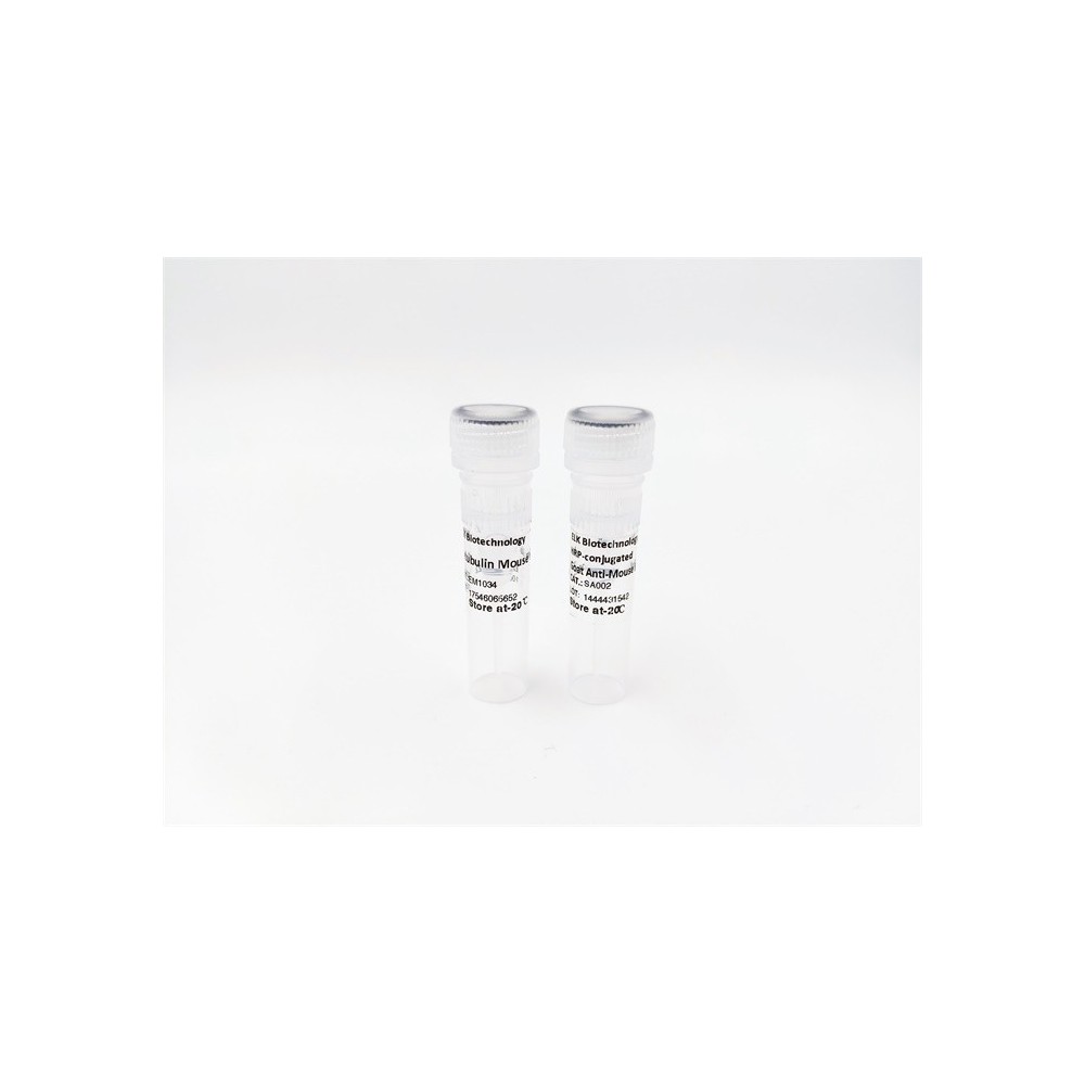

| ELK.No | ES4126 |
| Product name | CD166 rabbit pAb |
| Reactivity | Human;Mouse;Rat |
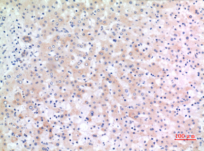
| Applications | IF;WB;ELISA |
| Other name | ALCAM; MEMD; CD166 antigen; Activated leukocyte cell adhesion molecule; CD166 |
| Size | 50μL |
| Unit price ($) | 148 |
| Human gene ID | 214 |
| Human Swiss-Prot | Q13740 |
| Source | Rabbit |
| Isotype | IgG |
| Target | CD166 |
| Fields | >>Cell adhesion molecules |
| Gene name | ALCAM |
| Protein name | CD166 antigen |
| Human gene link | View Human Gene |
| Human Swiss link | View Human Swiss-Prot |
| Mouse gene ID | 11658 |
| Mouse gene link | View Mouse Gene |
| Mouse Swiss-Prot | Q61490 |
| Mouse Swiss link | View Mouse Swiss-Prot |
| Rat gene ID | 79559 |
| Rat gene link | View Rat Gene |
| Rat Swiss-Prot | O35112 |
| Rat Swiss link | View Rat Swiss-Prot |
| Immunogen | The antiserum was produced against synthesized peptide derived from the C-terminal region of human ALCAM. AA range:481-530 |
| Specificity | CD166 Polyclonal Antibody detects endogenous levels of CD166 protein. |
| Formulation | Liquid in PBS containing 50% glycerol, 0.5% BSA and 0.02% sodium azide. |
| Clonality | Polyclonal |
| Dilution | IF: 1:50-200 Western Blot: 1/500 - 1/2000. ELISA: 1/20000. Not yet tested in other applications. |
| Purification | The antibody was affinity-purified from rabbit antiserum by affinity-chromatography using epitope-specific immunogen. |
| Concentration | 1 mg/ml |
| Storage stability | -20°C/1 year |
| Molecular Weight (Da) | |
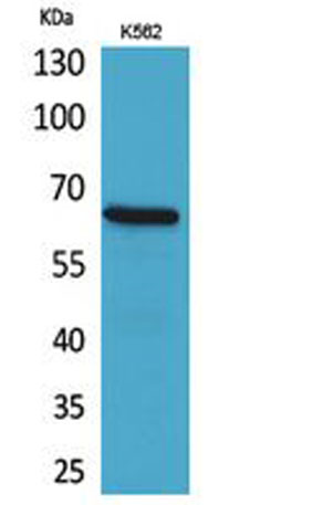
| Observed band (KD) | 65kD |
| Background | This gene encodes activated leukocyte cell adhesion molecule (ALCAM), also known as CD166 (cluster of differentiation 166), which is a member of a subfamily of immunoglobulin receptors with five immunoglobulin-like domains (VVC2C2C2) in the extracellular domain. This protein binds to T-cell differentiation antigene CD6, and is implicated in the processes of cell adhesion and migration. Multiple alternatively spliced transcript variants encoding different isoforms have been found. [provided by RefSeq, Aug 2011], |
| Function | domain:The CD6 binding site is located in the N-terminal Ig-like domain.,function:Cell adhesion molecule that binds to CD6. Involved in neurite extension by neurons via heterophilic and homophilic interactions. May play a role in the binding of T- and B-cells to activated leukocytes, as well as in interactions between cells of the nervous system.,similarity:Contains 2 Ig-like V-type (immunoglobulin-like) domains.,similarity:Contains 3 Ig-like C2-type (immunoglobulin-like) domains.,tissue specificity:Spleen, placenta, liver, and weakly in liver. Expressed by activated T-cells, B-cells, monocytes and thymic epithelial cells. Expressed by neurons in the brain. Restricted expression in tumor cell lines. Preferentially expressed in highly metastasizing melanoma cell lines., |
| Subcellular location | Cell membrane ; Single-pass type I membrane protein . Cell projection, axon . Cell projection, dendrite . Detected at the immunological synapse, i.e, at the contact zone between antigen-presenting dendritic cells and T-cells (PubMed:15294938, PubMed:16352806). Colocalizes with CD6 and the TCR/CD3 complex at the immunological synapse (PubMed:15294938). .; [Isoform 3]: Secreted . |
| Expression | Detected on hematopoietic stem cells derived from umbilical cord blood (PubMed:24740813). Detected on lymph vessel endothelial cells, skin and tonsil (PubMed:23169771). Detected on peripheral blood monocytes (PubMed:15048703). Detected on monocyte-derived dendritic cells (at protein level) (PubMed:16352806). Detected at low levels in spleen, placenta, liver (PubMed:9502422). Expressed by activated T-cells, B-cells, monocytes and thymic epithelial cells (PubMed:7760007). Isoform 1 and isoform 3 are detected in vein and artery endothelial cells, astrocytes, keratinocytes and artery smooth muscle cells (PubMed:15496415). Expressed by neurons in the brain. Restricted expression in tumor cell lines. Detected in highly metastasizing melanoma cell lines (PubMed:9502422). |

Use collapsible tabs for more detailed information that will help customers make a purchasing decision.
Ex: Shipping and return policies, size guides, and other common questions.