 Français
 English

| ELK.No | ES4131 |
| Product name | Jagged1 rabbit pAb |
| Reactivity | Human;Mouse;Rat |
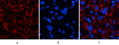
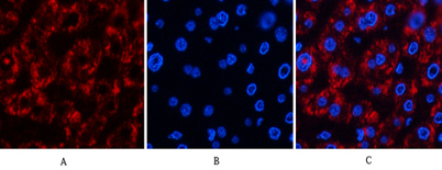
| Applications | IF;WB;IHC;ELISA |
| Other name | JAG1; JAGL1; Protein jagged-1; Jagged1; hJ1; CD339 |
| Size | 50μL |
| Unit price ($) | 148 |
| Human gene ID | 182 |
| Human Swiss-Prot | P78504 |
| Source | Rabbit |
| Isotype | IgG |
| Target | Jagged1 |
| Fields | >>Endocrine resistance;>>Notch signaling pathway;>>Apelin signaling pathway;>>Th1 and Th2 cell differentiation;>>TNF signaling pathway;>>Human papillomavirus infection;>>Pathways in cancer;>>Chemical carcinogenesis - receptor activation;>>Breast cancer |
| Gene name | JAG1 |
| Protein name | Protein jagged-1 |
| Human gene link | View Human Gene |
| Human Swiss link | View Human Swiss-Prot |
| Mouse gene ID | 16449 |
| Mouse gene link | View Mouse Gene |
| Mouse Swiss-Prot | Q9QXX0 |
| Mouse Swiss link | View Mouse Swiss-Prot |
| Rat gene ID | 29146 |
| Rat gene link | View Rat Gene |
| Rat Swiss-Prot | Q63722 |
| Rat Swiss link | View Rat Swiss-Prot |
| Immunogen | The antiserum was produced against synthesized peptide derived from the Internal region of human JAG1. AA range:981-1030 |
| Specificity | Jagged1 Polyclonal Antibody detects endogenous levels of Jagged1 protein. |
| Formulation | Liquid in PBS containing 50% glycerol, 0.5% BSA and 0.02% sodium azide. |
| Clonality | Polyclonal |
| Dilution | IF: 1:50-200 Western Blot: 1/500 - 1/2000. IHC-p: 1/100-1/300. ELISA: 1/20000. Not yet tested in other applications. |
| Purification | The antibody was affinity-purified from rabbit antiserum by affinity-chromatography using epitope-specific immunogen. |
| Concentration | 1 mg/ml |
| Storage stability | -20°C/1 year |
| Molecular Weight (Da) | |
| Observed band (KD) | 140kD |
| Background | The jagged 1 protein encoded by JAG1 is the human homolog of the Drosophilia jagged protein. Human jagged 1 is the ligand for the receptor notch 1, the latter a human homolog of the Drosophilia jagged receptor notch. Mutations that alter the jagged 1 protein cause Alagille syndrome. Jagged 1 signalling through notch 1 has also been shown to play a role in hematopoiesis. [provided by RefSeq, Jul 2008], |
| Function | developmental stage:Expressed in 32-52 days embryos in the distal cardiac outflow tract and pulmonary artery, major arteries, portal vein, optic vesicle, otocyst, branchial arches, metanephros, pancreas, mesocardium, around the major bronchial branches, and in the neural tube.,disease:Defects in JAG1 are a cause of tetralogy of Fallot (TOF) [MIM:187500]. TOF is a congenital heart anomaly which consists of pulmonary stenosis, ventricular septal defect, dextroposition of the aorta (aorta is on the right side instead of the left) and hypertrophy of the right ventricle. This condition results in a blue baby at birth due to inadequate oxygenation. Surgical correction is emergent.,disease:Defects in JAG1 are the cause of Alagille syndrome type 1 (ALGS1) [MIM:118450]. Alagille syndrome is an autosomal dominant multisystem disorder defined clinically by hepatic bile duct paucity and cholestasis |
| Subcellular location | Membrane; Single-pass type I membrane protein. |
| Expression | Widely expressed in adult and fetal tissues. In cervix epithelium expressed in undifferentiated subcolumnar reserve cells and squamous metaplasia. Expression is up-regulated in cervical squamous cell carcinoma. Expressed in bone marrow cell line HS-27a which supports the long-term maintenance of immature progenitor cells. |


Use collapsible tabs for more detailed information that will help customers make a purchasing decision.
Ex: Shipping and return policies, size guides, and other common questions.