 Français
 English
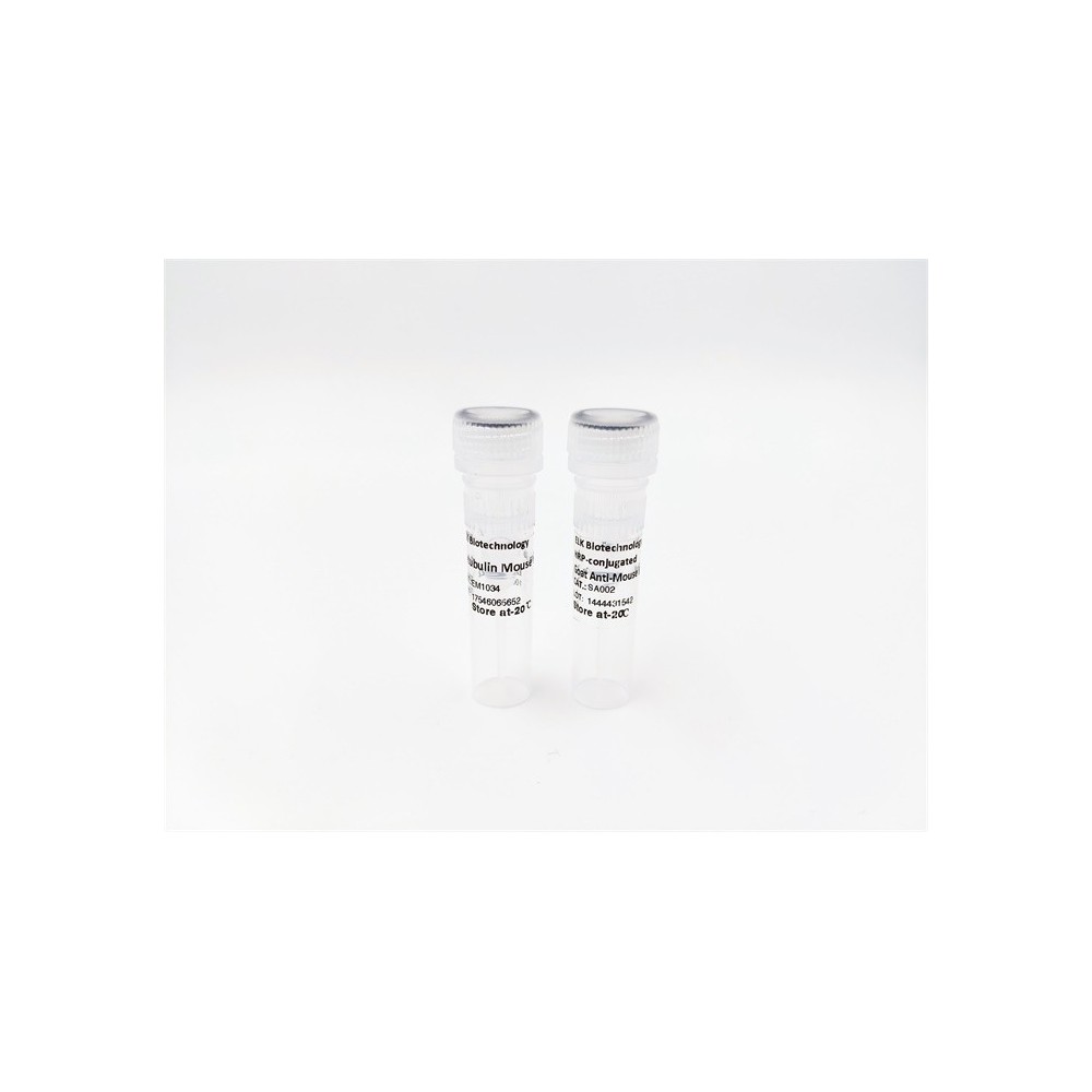

| ELK.No | ES4134 |
| Product name | RANKL rabbit pAb |
| Reactivity | Human;Mouse;Rat |
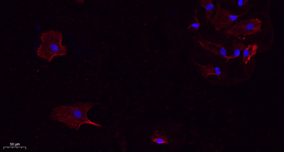
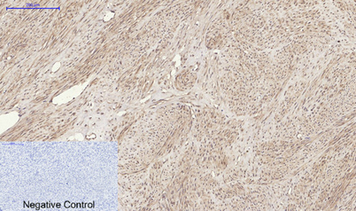
| Applications | WB;IHC;IF;ELISA |
| Other name | TNFSF11; OPGL; RANKL; TRANCE; Tumor necrosis factor ligand superfamily member 11; Osteoclast differentiation factor; ODF; Osteoprotegerin ligand; OPGLReceptor activator of nuclear factor kappa-B ligand; RANKL; TNF-related activation-induced cytokine; TRANCE; CD254 |
| Size | 50μL |
| Unit price ($) | 148 |
| Human gene ID | 8600 |
| Human Swiss-Prot | O14788 |
| Source | Rabbit |
| Isotype | IgG |
| Target | RANKL |
| Fields | >>Cytokine-cytokine receptor interaction;>>NF-kappa B signaling pathway;>>Osteoclast differentiation;>>Prolactin signaling pathway;>>Parathyroid hormone synthesis, secretion and action;>>Chemical carcinogenesis - receptor activation;>>Breast cancer;>>Rheumatoid arthritis |
| Gene name | TNFSF11 |
| Protein name | Tumor necrosis factor ligand superfamily member 11 |
| Human gene link | View Human Gene |
| Human Swiss link | View Human Swiss-Prot |
| Mouse gene ID | 21943 |
| Mouse gene link | View Mouse Gene |
| Mouse Swiss-Prot | O35235 |
| Mouse Swiss link | View Mouse Swiss-Prot |
| Rat gene ID | 117516 |
| Rat gene link | View Rat Gene |
| Rat Swiss-Prot | Q9ESE2 |
| Rat Swiss link | View Rat Swiss-Prot |
| Immunogen | The antiserum was produced against synthesized peptide derived from the C-terminal region of human TNFSF11. AA range:268-317 |
| Specificity | RANKL Polyclonal Antibody detects endogenous levels of RANKL protein. |
| Formulation | Liquid in PBS containing 50% glycerol, 0.5% BSA and 0.02% sodium azide. |
| Clonality | Polyclonal |
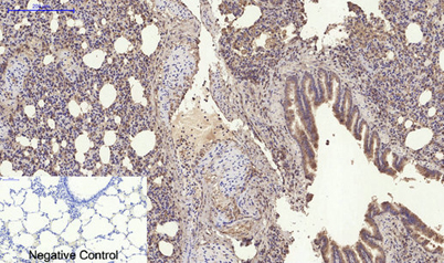
| Dilution | Western Blot: 1/500 - 1/2000. IHC-p: 1:100-300 ELISA: 1/20000. IF 1:100-300 Not yet tested in other applications. |
| Purification | The antibody was affinity-purified from rabbit antiserum by affinity-chromatography using epitope-specific immunogen. |
| Concentration | 1 mg/ml |
| Storage stability | -20°C/1 year |
| Molecular Weight (Da) | |
| Observed band (KD) | 35kD |
| Background | This gene encodes a member of the tumor necrosis factor (TNF) cytokine family which is a ligand for osteoprotegerin and functions as a key factor for osteoclast differentiation and activation. This protein was shown to be a dentritic cell survival factor and is involved in the regulation of T cell-dependent immune response. T cell activation was reported to induce expression of this gene and lead to an increase of osteoclastogenesis and bone loss. This protein was shown to activate antiapoptotic kinase AKT/PKB through a signaling complex involving SRC kinase and tumor necrosis factor receptor-associated factor (TRAF) 6, which indicated this protein may have a role in the regulation of cell apoptosis. Targeted disruption of the related gene in mice led to severe osteopetrosis and a lack of osteoclasts. The deficient mice exhibited defects in early differentiation of T and B ly |
| Function | disease:Defects in TNFSF11 are the cause of osteopetrosis autosomal recessive type 2 (OPTB2) [MIM:259710]; also known as osteoclast-poor osteopetrosis. Osteopetrosis is a rare genetic disease characterized by abnormally dense bone, due to defective resorption of immature bone. The disorder occurs in two forms: a severe autosomal recessive form occurring in utero, infancy, or childhood, and a benign autosomal dominant form occurring in adolescence or adulthood. Autosomal recessive osteopetrosis is usually associated with normal or elevated amount of non-functional osteoclasts. OPTB2 is characterized by paucity of osteoclasts, suggesting a molecular defect in osteoclast development.,function:Cytokine that binds to TNFRSF11B/OPG and to TNFRSF11A/RANK. Osteoclast differentiation and activation factor. Augments the ability of dendritic cells to stimulate naive T-cell proliferation. May be an |
| Subcellular location | [Isoform 1]: Cell membrane; Single-pass type II membrane protein.; [Isoform 3]: Cell membrane; Single-pass type II membrane protein.; [Isoform 2]: Cytoplasm .; [Tumor necrosis factor ligand superfamily member 11, soluble form]: Secreted . |
| Expression | Highest in the peripheral lymph nodes, weak in spleen, peripheral blood Leukocytes, bone marrow, heart, placenta, skeletal muscle, stomach and thyroid. |

Use collapsible tabs for more detailed information that will help customers make a purchasing decision.
Ex: Shipping and return policies, size guides, and other common questions.