 Français
 English
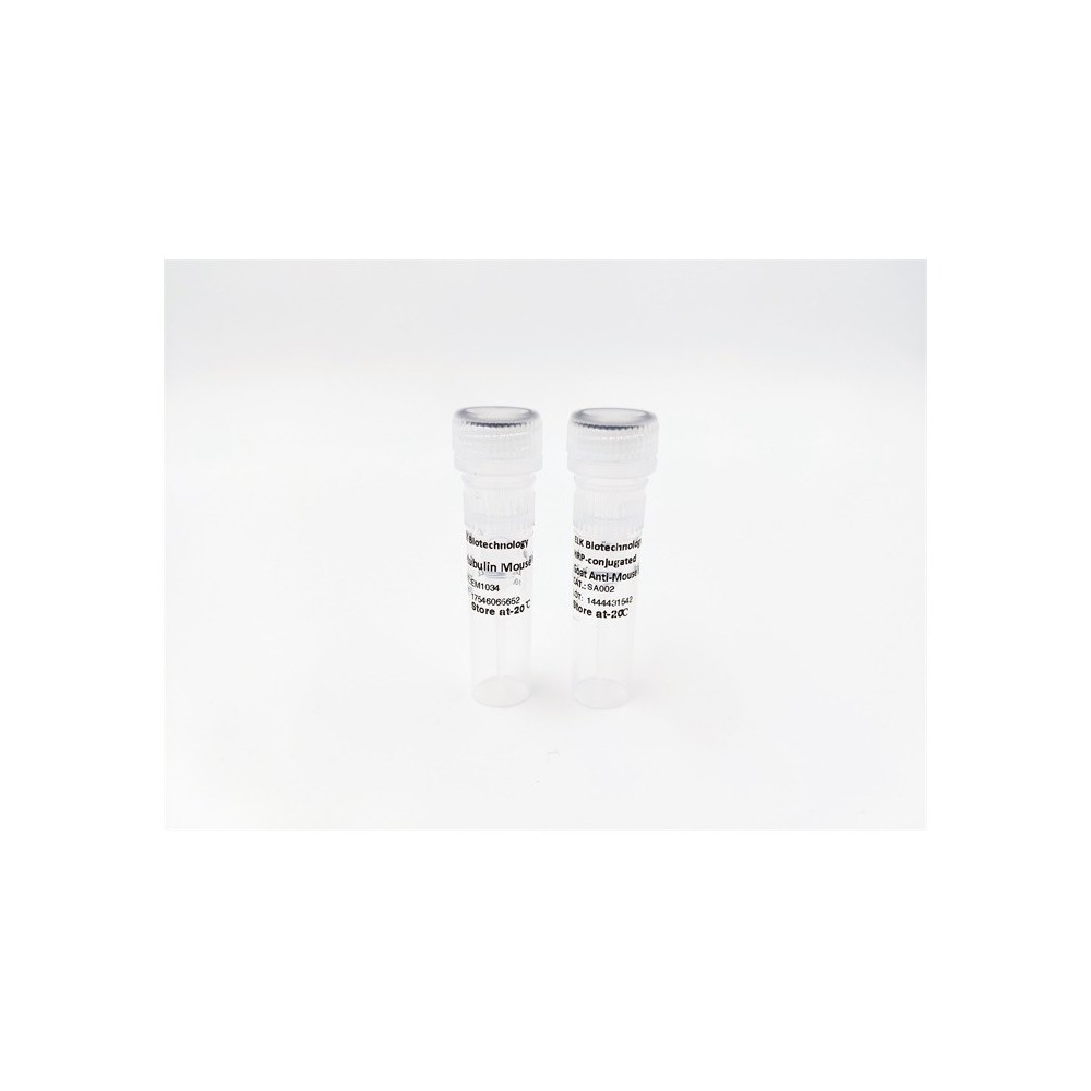

| ELK.No | ES4288 |
| Product name | ABCG2 rabbit pAb |
| Reactivity | Human;Rat;Mouse; |
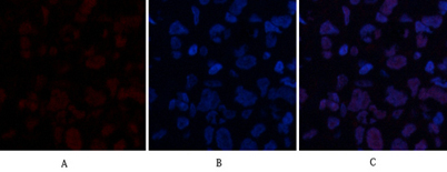
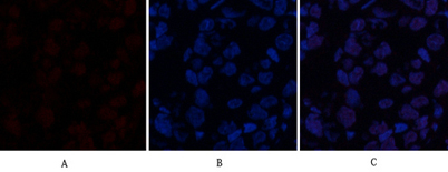
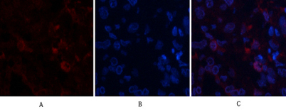
| Applications | IF;WB;IHC;ELISA |
| Other name | ABCG2; ABCP; BCRP; BCRP1; MXR; ATP-binding cassette sub-family G member 2; Breast cancer resistance protein; CDw338; Mitoxantrone resistance-associated protein; Placenta-specific ATP-binding cassette transporter; CD338 |
| Size | 50μL |
| Unit price ($) | 148 |
| Human gene ID | 9429 |
| Human Swiss-Prot | Q9UNQ0 |
| Source | Rabbit |
| Isotype | IgG |
| Target | ABCG2 |
| Fields | >>Antifolate resistance;>>ABC transporters;>>Bile secretion |
| Gene name | ABCG2 |
| Protein name | ATP-binding cassette sub-family G member 2 |
| Human gene link | View Human Gene |
| Human Swiss link | View Human Swiss-Prot |
| Mouse gene ID | |
| Mouse gene link | |
| Mouse Swiss-Prot | Q7TMS5 |
| Mouse Swiss link | View Mouse Swiss-Prot |
| Rat gene ID | |
| Rat gene link | |
| Rat Swiss-Prot | |
| Rat Swiss link | |
| Immunogen | The antiserum was produced against synthesized peptide derived from the Internal region of human ABCG2. AA range:461-510 |
| Specificity | ABCG2 Polyclonal Antibody detects endogenous levels of ABCG2 protein. |
| Formulation | Liquid in PBS containing 50% glycerol, 0.5% BSA and 0.02% sodium azide. |
| Clonality | Polyclonal |
| Dilution | IF: 1:50-200 Western Blot: 1/500 - 1/2000. IHC-p: 1:100-1:300. ELISA: 1/10000. Not yet tested in other applications. |
| Purification | The antibody was affinity-purified from rabbit antiserum by affinity-chromatography using epitope-specific immunogen. |
| Concentration | 1 mg/ml |
| Storage stability | -20°C/1 year |
| Molecular Weight (Da) | |
| Observed band (KD) | 75kD |
| Background | The membrane-associated protein encoded by this gene is included in the superfamily of ATP-binding cassette (ABC) transporters. ABC proteins transport various molecules across extra- and intra-cellular membranes. ABC genes are divided into seven distinct subfamilies (ABC1, MDR/TAP, MRP, ALD, OABP, GCN20, White). This protein is a member of the White subfamily. Alternatively referred to as a breast cancer resistance protein, this protein functions as a xenobiotic transporter which may play a major role in multi-drug resistance. It likely serves as a cellular defense mechanism in response to mitoxantrone and anthracycline exposure. Significant expression of this protein has been observed in the placenta, which may suggest a potential role for this molecule in placenta tissue. Multiple transcript variants encoding different isoforms have been found for this gene. |
| Function | function:Xenobiotic transporter that may play an important role in the exclusion of xenobiotics from the brain. May be involved in brain-to-blood efflux. Appears to play a major role in the multidrug resistance phenotype of several cancer cell lines. When overexpressed, the transfected cells become resistant to mitoxantrone, daunorubicin and doxorubicin, display diminished intracellular accumulation of daunorubicin, and manifest an ATP-dependent increase in the efflux of rhodamine 123.,induction:Up-regulated in brain tumors.,PTM:Glycosylation-deficient ABCG2 is normally expressed and functional.,similarity:Belongs to the ABC transporter family. ABCG (White) subfamily.,similarity:Contains 1 ABC transmembrane type-2 domain.,similarity:Contains 1 ABC transporter domain.,subunit:Monomer or homodimer; disulfide-linked.,tissue specificity:Highly expressed in placenta. Low expression in small i |
| Subcellular location | Cell membrane ; Multi-pass membrane protein . Apical cell membrane ; Multi-pass membrane protein . Mitochondrion membrane ; Multi-pass membrane protein . Enriched in membrane lipid rafts. . |
| Expression | Highly expressed in placenta (PubMed:9850061). Low expression in small intestine, liver and colon (PubMed:9861027). Expressed in brain (at protein level) (PubMed:12958161). |

Use collapsible tabs for more detailed information that will help customers make a purchasing decision.
Ex: Shipping and return policies, size guides, and other common questions.