 Français
 English
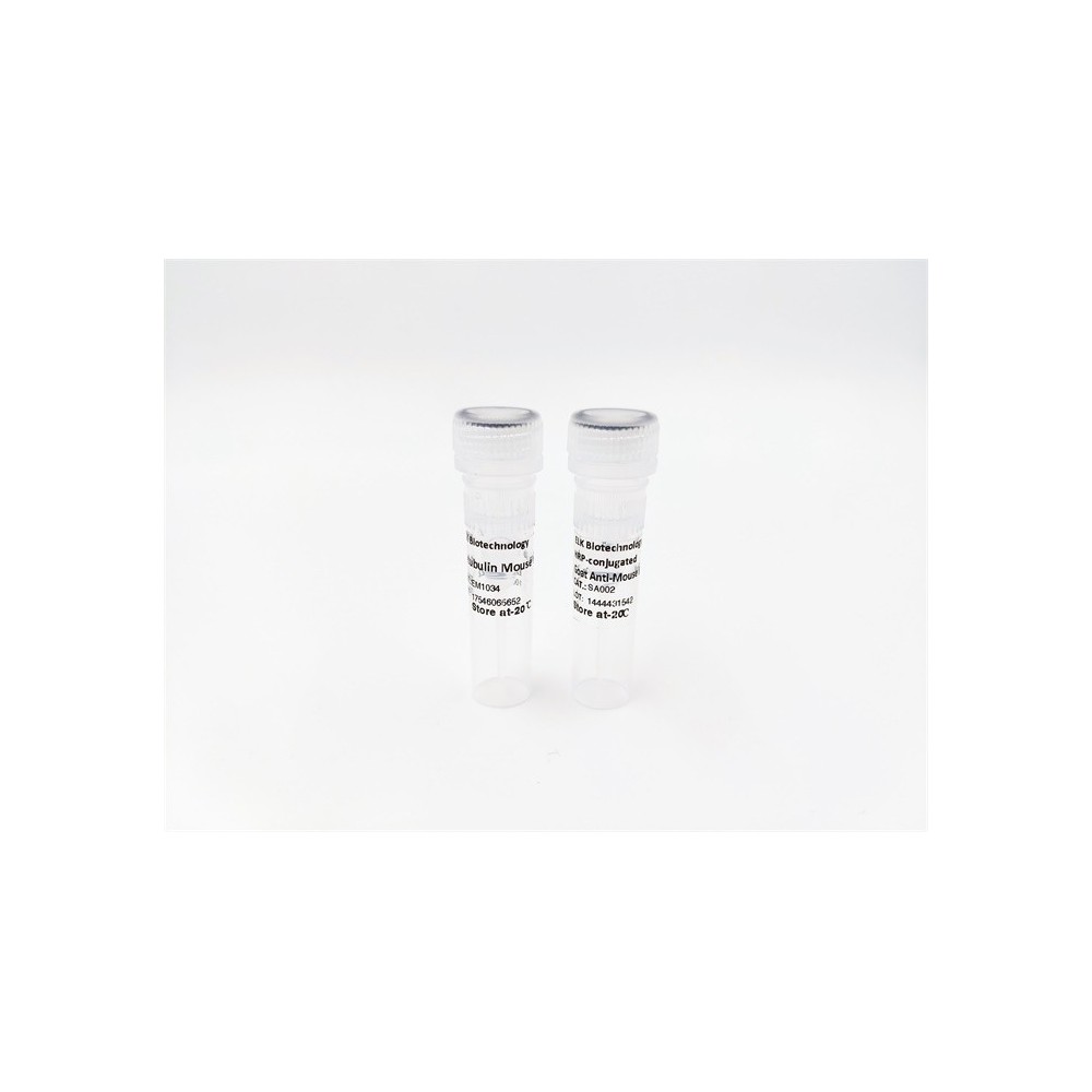

| ELK.No | ES4326 |
| Product name | LIR-1 rabbit pAb |
| Reactivity | Human;Rat;Mouse; |
| Applications | WB;IHC |
| Other name | LILRB1; ILT2; LIR1; MIR7; Leukocyte immunoglobulin-like receptor subfamily B member 1; LIR-1; Leukocyte immunoglobulin-like receptor 1; CD85 antigen-like family member J; Immunoglobulin-like transcript 2; ILT-2; Monocyte/macrophage immunoglobulin-like receptor 7; MIR-7; CD85j |
| Size | 100μL |
| Unit price ($) | 248 |
| Human gene ID | 10859 |
| Human Swiss-Prot | Q8NHL6 |
| Source | Rabbit |
| Isotype | IgG |
| Target | LIR-1 |
| Fields | >>Osteoclast differentiation;>>B cell receptor signaling pathway |
| Gene name | LILRB1 |
| Protein name | Leukocyte immunoglobulin-like receptor subfamily B member 1 |
| Human gene link | View Human Gene |
| Human Swiss link | View Human Swiss-Prot |
| Mouse gene ID | |
| Mouse gene link | |
| Mouse Swiss-Prot | |
| Mouse Swiss link | |
| Rat gene ID | |
| Rat gene link | |
| Rat Swiss-Prot | |
| Rat Swiss link | |
| Immunogen | The antiserum was produced against synthesized peptide derived from the N-terminal region of human LILRB1. AA range:21-70 |
| Specificity | LIR-1 Polyclonal Antibody detects endogenous levels of LIR-1 protein. |
| Formulation | Liquid in PBS containing 50% glycerol, 0.5% BSA and 0.02% sodium azide. |
| Clonality | Polyclonal |
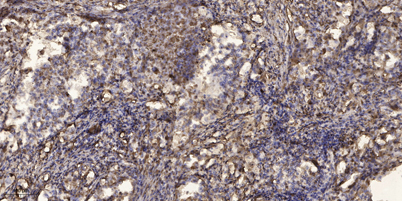
| Dilution | WB 1:500-2000;IHC-p 1:50-300 |
| Purification | The antibody was affinity-purified from rabbit antiserum by affinity-chromatography using epitope-specific immunogen. |
| Concentration | 1 mg/ml |
| Storage stability | -20°C/1 year |
| Molecular Weight (Da) | |
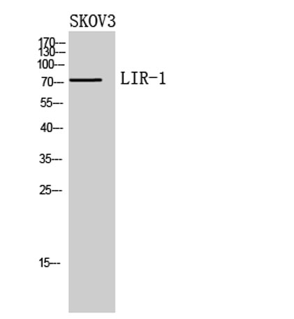
| Observed band (KD) | 72kD |
| Background | This gene is a member of the leukocyte immunoglobulin-like receptor (LIR) family, which is found in a gene cluster at chromosomal region 19q13.4. The encoded protein belongs to the subfamily B class of LIR receptors which contain two or four extracellular immunoglobulin domains, a transmembrane domain, and two to four cytoplasmic immunoreceptor tyrosine-based inhibitory motifs (ITIMs). The receptor is expressed on immune cells where it binds to MHC class I molecules on antigen-presenting cells and transduces a negative signal that inhibits stimulation of an immune response. It is thought to control inflammatory responses and cytotoxicity to help focus the immune response and limit autoreactivity. Multiple transcript variants encoding different isoforms have been found for this gene. [provided by RefSeq, Jul 2008], |
| Function | domain:Contains 4 copies of a cytoplasmic motif that is referred to as the immunoreceptor tyrosine-based inhibitor motif (ITIM). This motif is involved in modulation of cellular responses. The phosphorylated ITIM motif can bind the SH2 domain of several SH2-containing phosphatases.,function:Receptor for class I MHC antigens. Recognizes a broad spectrum of HLA-A, HLA-B, HLA-C and HLA-G alleles. Receptor for H301/UL18, a human cytomegalovirus class I MHC homolog. Ligand binding results in inhibitory signals and down-regulation of the immune response. Engagement of LILRB1 present on natural killer cells or T-cells by class I MHC molecules protects the target cells from lysis. Interaction with HLA-B or HLA-E leads to inhibition of the signal triggered by FCER1A and inhibits serotonin release. Inhibits FCGR1A-mediated phosphorylation of cellular proteins and mobilization of intracellular calc |
| Subcellular location | Cell membrane ; Single-pass type I membrane protein .; [Isoform 5]: Secreted . |
| Expression | Expressed in B cells, monocytes and various dendritic cell (DC) subsets including myeloid, plasmacytoid and tolerogenic DCs (at protein level) (PubMed:20448110, PubMed:9285411, PubMed:9842885, PubMed:24453251). Expressed in decidual macrophages (at protein level) (PubMed:19304799). Expressed in decidual NK cells (at protein level) (PubMed:29262349). |

Use collapsible tabs for more detailed information that will help customers make a purchasing decision.
Ex: Shipping and return policies, size guides, and other common questions.