 Français
 English

| ELK.No | ES4329 |
| Product name | CD98 rabbit pAb |
| Reactivity | Human;Mouse;Rat |
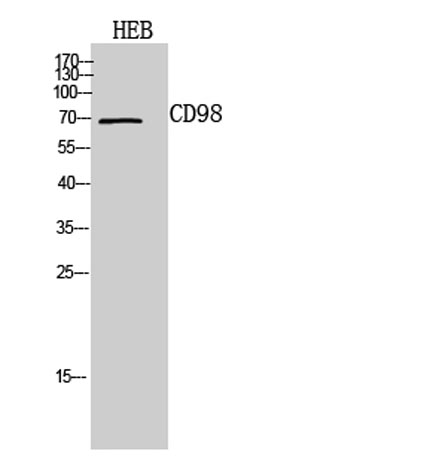
| Applications | WB;ELISA |
| Other name | SLC3A2; MDU1; 4F2 cell-surface antigen heavy chain; 4F2hc; 4F2 heavy chain antigen; Lymphocyte activation antigen 4F2 large subunit; CD98 |
| Size | 100μL |
| Unit price ($) | 248 |
| Human gene ID | 6520 |
| Human Swiss-Prot | P08195 |
| Source | Rabbit |
| Isotype | IgG |
| Target | CD98 |
| Fields | >>mTOR signaling pathway;>>Ferroptosis;>>Protein digestion and absorption |
| Gene name | SLC3A2 |
| Protein name | 4F2 cell-surface antigen heavy chain |
| Human gene link | View Human Gene |
| Human Swiss link | View Human Swiss-Prot |
| Mouse gene ID | 17254 |
| Mouse gene link | View Mouse Gene |
| Mouse Swiss-Prot | P10852 |
| Mouse Swiss link | View Mouse Swiss-Prot |
| Rat gene ID | 50567 |
| Rat gene link | View Rat Gene |
| Rat Swiss-Prot | Q794F9 |
| Rat Swiss link | View Rat Swiss-Prot |
| Immunogen | The antiserum was produced against synthesized peptide derived from the C-terminal region of human SLC3A2. AA range:491-540 |
| Specificity | CD98 Polyclonal Antibody detects endogenous levels of CD98 protein. |
| Formulation | Liquid in PBS containing 50% glycerol, 0.5% BSA and 0.02% sodium azide. |
| Clonality | Polyclonal |
| Dilution | Western Blot: 1/500 - 1/2000. ELISA: 1/10000. Not yet tested in other applications. |
| Purification | The antibody was affinity-purified from rabbit antiserum by affinity-chromatography using epitope-specific immunogen. |
| Concentration | 1 mg/ml |
| Storage stability | -20°C/1 year |
| Molecular Weight (Da) | |
| Observed band (KD) | 69kD |
| Background | This gene is a member of the solute carrier family and encodes a cell surface, transmembrane protein. The protein exists as the heavy chain of a heterodimer, covalently bound through di-sulfide bonds to one of several possible light chains. The encoded transporter plays a role in regulation of intracellular calcium levels and transports L-type amino acids. Alternatively spliced transcript variants, encoding different isoforms, have been characterized. [provided by RefSeq, Nov 2010], |
| Function | function:Required for the function of light chain amino-acid transporters. Involved in sodium-independent, high-affinity transport of large neutral amino acids such as phenylalanine, tyrosine, leucine, arginine and tryptophan. Involved in guiding and targeting of LAT1 and LAT2 to the plasma membrane. When associated with SLC7A6 or SLC7A7 acts as an arginine/glutamine exchanger, following an antiport mechanism for amino acid transport, influencing arginine release in exchange for extracellular amino acids. Plays a role in nitric oxide synthesis in human umbilical vein endothelial cells (HUVECs) via transport of L-arginine. Required for normal and neoplastic cell growth. When associated with SLC7A5/LAT1, is also involved in the transport of L-DOPA across the blood-brain barrier, and that of thyroid hormones triiodothyronine (T3) and thyroxine (T4) across the cell membrane in tissues such a |
| Subcellular location | Apical cell membrane . Cell membrane ; Single-pass type II membrane protein . Cell junction . Lysosome membrane . Melanosome . Localized at the plasma membrane when associated with SLC7A5/LAT1 or SLC7A8/LAT2 (PubMed:9751058, PubMed:11311135). Localized to the apical membrane of placental syncytiotrophoblastic cells (PubMed:11742812). Recruited to lysosomes by LAPTM4B (PubMed:25998567). Identified by mass spectrometry in melanosome fractions from stage I to stage IV (PubMed:17081065). Located selectively at cell-cell adhesion sites (By similarity). Colocalized with SLC7A8/LAT2 at the basolateral membrane of kidney proximal tubules and small intestine epithelia. Expressed in both luminal and abluminal membranes of brain capillary endothelial cells (By similarity). . |
| Expression | Expressed ubiquitously in all tissues tested with highest levels detected in kidney, placenta and testis and weakest level in thymus. During gestation, expression in the placenta was significantly stronger at full-term than at the mid-trimester stage. Expressed in HUVECS and at low levels in resting peripheral blood T-lymphocytes and quiescent fibroblasts. Also expressed in fetal liver and in the astrocytic process of primary astrocytic gliomas. Expressed in retinal endothelial cells and in the intestinal epithelial cell line C2BBe1. |

Use collapsible tabs for more detailed information that will help customers make a purchasing decision.
Ex: Shipping and return policies, size guides, and other common questions.