 Français
 English
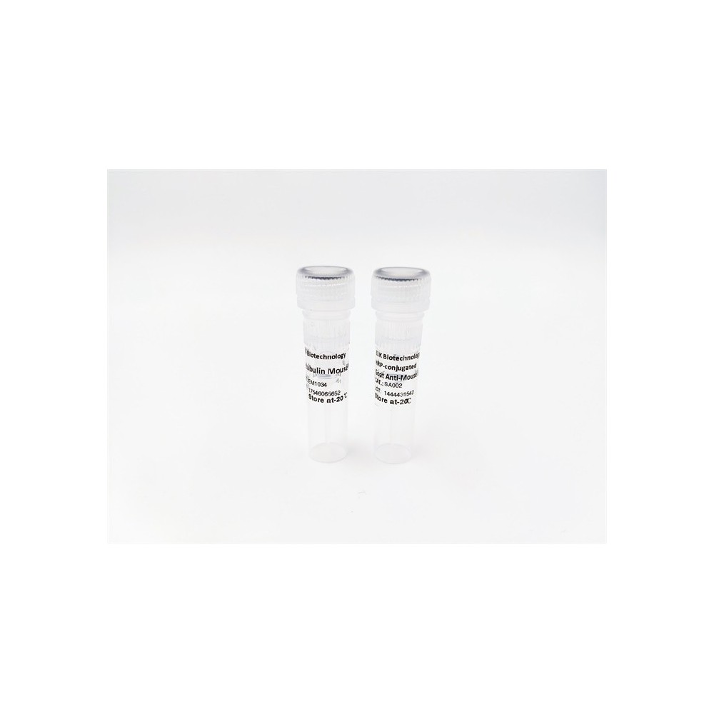

| ELK.No | ES4330 |
| Product name | CD101 rabbit pAb |
| Reactivity | Human;Rat;Mouse; |
| Applications | WB;ELISA |
| Other name | CD101; EWI101; IGSF2; V7; Immunoglobulin superfamily member 2; IgSF2; Cell surface glycoprotein V7; Glu-Trp-Ile EWI motif-containing protein 101; EWI-101; CD101 |
| Size | 100μL |
| Unit price ($) | 248 |
| Human gene ID | 9398 |
| Human Swiss-Prot | Q93033 |
| Source | Rabbit |
| Isotype | IgG |
| Target | CD101 |
| Fields | |
| Gene name | CD101 |
| Protein name | Immunoglobulin superfamily member 2 |
| Human gene link | View Human Gene |
| Human Swiss link | View Human Swiss-Prot |
| Mouse gene ID | |
| Mouse gene link | |
| Mouse Swiss-Prot | A8E0Y8 |
| Mouse Swiss link | View Mouse Swiss-Prot |
| Rat gene ID | |
| Rat gene link | |
| Rat Swiss-Prot | |
| Rat Swiss link | |
| Immunogen | The antiserum was produced against synthesized peptide derived from the Internal region of human CD101. AA range:731-780 |
| Specificity | CD101 Polyclonal Antibody detects endogenous levels of CD101 protein. |
| Formulation | Liquid in PBS containing 50% glycerol, 0.5% BSA and 0.02% sodium azide. |
| Clonality | Polyclonal |
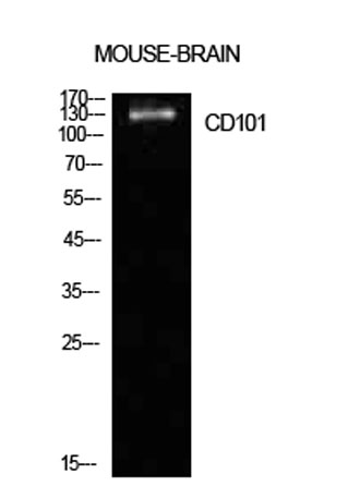
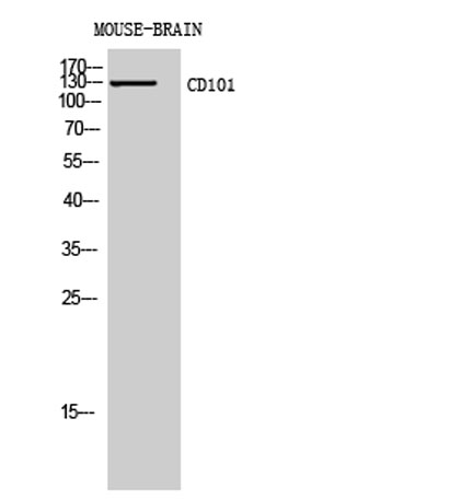
| Dilution | Western Blot: 1/500 - 1/2000. ELISA: 1/10000. Not yet tested in other applications. |
| Purification | The antibody was affinity-purified from rabbit antiserum by affinity-chromatography using epitope-specific immunogen. |
| Concentration | 1 mg/ml |
| Storage stability | -20°C/1 year |
| Molecular Weight (Da) | |
| Observed band (KD) | 130kD |
| Background | function:Plays a role as inhibitor of T-cells proliferation induced by CD3. Inhibits expression of IL2RA on activated T-cells and secretion of IL2. Inhibits tyrosine kinases that are required for IL2 production and cellular proliferation. Inhibits phospholipase C-gamma-1/PLCG1 phosphorylation and subsequent CD3-induced changes in intracellular free calcium. Prevents nuclear translocation of nuclear factor of activated T-cell to the nucleus. Plays a role in the inhibition of T-cell proliferation via IL10 secretion by cutaneous dendritic cells. May be a marker of CD4(+) CD56(+) leukemic tumor cells.,PTM:N-glycosylated.,similarity:Contains 7 Ig-like C2-type (immunoglobulin-like) domains.,tissue specificity:Expressed in lung, thymus and small intestine. Detected in cutaneous dendritic cells, activated T-cells, monocytes and granulocytes as well as with epithelial cells with dendritic morphology. Expressed in some leukemic cells, the CD4(+) CD56(+) blastic tumor cells, as well as in Langerhans cells from LCH (Langerhans cell histiocytosis) patients., |
| Function | function:Plays a role as inhibitor of T-cells proliferation induced by CD3. Inhibits expression of IL2RA on activated T-cells and secretion of IL2. Inhibits tyrosine kinases that are required for IL2 production and cellular proliferation. Inhibits phospholipase C-gamma-1/PLCG1 phosphorylation and subsequent CD3-induced changes in intracellular free calcium. Prevents nuclear translocation of nuclear factor of activated T-cell to the nucleus. Plays a role in the inhibition of T-cell proliferation via IL10 secretion by cutaneous dendritic cells. May be a marker of CD4(+) CD56(+) leukemic tumor cells.,PTM:N-glycosylated.,similarity:Contains 7 Ig-like C2-type (immunoglobulin-like) domains.,tissue specificity:Expressed in lung, thymus and small intestine. Detected in cutaneous dendritic cells, activated T-cells, monocytes and granulocytes as well as with epithelial cells with dendritic morphol |
| Subcellular location | Membrane ; Single-pass type I membrane protein . |
| Expression | Expressed in lung, thymus and small intestine. Detected in cutaneous dendritic cells, activated T-cells, monocytes and granulocytes as well as with epithelial cells with dendritic morphology. Expressed in some leukemic cells, the CD4(+) CD56(+) blastic tumor cells, as well as in Langerhans cells from LCH (Langerhans cell histiocytosis) patients. |

Use collapsible tabs for more detailed information that will help customers make a purchasing decision.
Ex: Shipping and return policies, size guides, and other common questions.