 Français
 English
EphA2/3/4 (phospho Tyr588/596) rabbit pAb
AO-06-ES5072-100

| ELK.No | ES5072 |
| Product name | EphA2/3/4 (phospho Tyr588/596) rabbit pAb |
| Reactivity | Human;Mouse;Rat |
| Applications | WB;IF;ELISA |
| Other name | EPHA2; ECK; Ephrin type-A receptor 2; Epithelial cell kinase; Tyrosine-protein kinase receptor ECK; EPHA3; ETK; ETK1; HEK; TYRO4; Ephrin type-A receptor 3; EPH-like kinase 4; EK4; hEK4; HEK; Human embryo kinase; Tyrosine-protein kinase TYRO |
| Size | 100μL |
| Unit price ($) | 248 |
| Human gene ID | 1969/2042/2043 |
| Human Swiss-Prot | P29317/P29320/P54764 |
| Source | Rabbit |
| Isotype | IgG |
| Target | EphA2/3/4 |
| Fields | >>MAPK signaling pathway;>>Ras signaling pathway;>>Rap1 signaling pathway;>>PI3K-Akt signaling pathway;>>Axon guidance |
| Gene name | EPHA2/3/4 |
| Protein name | Ephrin type-A receptor 2/3/4 |
| Human gene link | View Human Gene |
| Human Swiss link | View Human Swiss-Prot |
| Mouse gene ID | 13836/13838 |
| Mouse gene link | View Mouse Gene |
| Mouse Swiss-Prot | |
| Mouse Swiss link | |
| Rat gene ID | 29210 |
| Rat gene link | View Rat Gene |
| Rat Swiss-Prot | O08680 |
| Rat Swiss link | View Rat Swiss-Prot |
| Immunogen | The antiserum was produced against synthesized peptide derived from human EPHA2/3 around the phosphorylation site of Tyr588/596. AA range:556-605 |
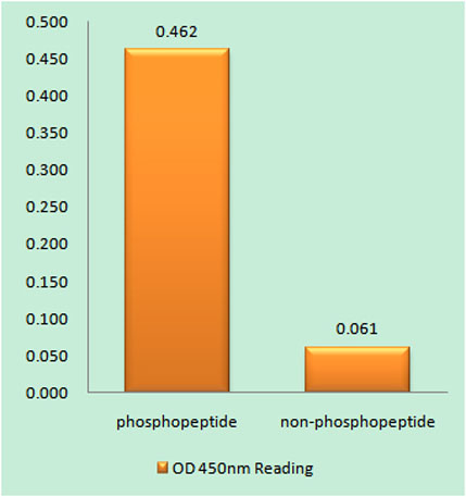
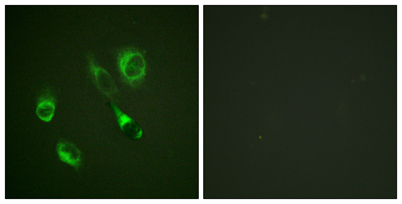
| Specificity | Phospho-EphA2/3/4 (Y588/596) Polyclonal Antibody detects endogenous levels of EphA2/3/4 protein only when phosphorylated at Y588/596. |
| Formulation | Liquid in PBS containing 50% glycerol, 0.5% BSA and 0.02% sodium azide. |
| Clonality | Polyclonal |
| Dilution | Western Blot: 1/500 - 1/2000. Immunofluorescence: 1/200 - 1/1000. ELISA: 1/20000. Not yet tested in other applications. |
| Purification | The antibody was affinity-purified from rabbit antiserum by affinity-chromatography using epitope-specific immunogen. |
| Concentration | 1 mg/ml |
| Storage stability | -20°C/1 year |
| Molecular Weight (Da) | |
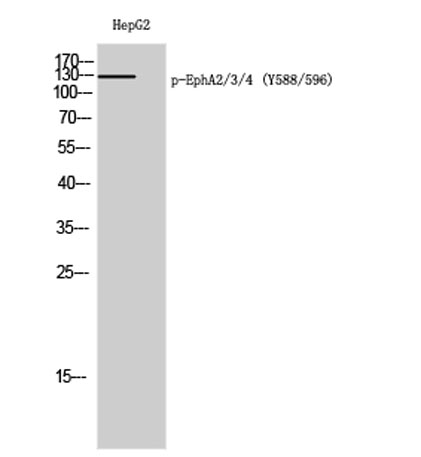
| Observed band (KD) | 130kD |
| Background | This gene belongs to the ephrin receptor subfamily of the protein-tyrosine kinase family. EPH and EPH-related receptors have been implicated in mediating developmental events, particularly in the nervous system. Receptors in the EPH subfamily typically have a single kinase domain and an extracellular region containing a Cys-rich domain and 2 fibronectin type III repeats. The ephrin receptors are divided into 2 groups based on the similarity of their extracellular domain sequences and their affinities for binding ephrin-A and ephrin-B ligands. This gene encodes a protein that binds ephrin-A ligands. Mutations in this gene are the cause of certain genetically-related cataract disorders.[provided by RefSeq, May 2010], |
| Function | catalytic activity:ATP + a [protein]-L-tyrosine = ADP + a [protein]-L-tyrosine phosphate.,function:Receptor for members of the ephrin-A family. Binds to ephrin-A1, -A3, -A4 and -A5.,similarity:Belongs to the protein kinase superfamily. Tyr protein kinase family. Ephrin receptor subfamily.,similarity:Contains 1 protein kinase domain.,similarity:Contains 1 SAM (sterile alpha motif) domain.,similarity:Contains 2 fibronectin type-III domains.,subunit:Interacts with SLA (By similarity). Interacts with INPPL1/SHIP2.,tissue specificity:Expressed most highly in tissues that contain a high proportion of epithelial cells, e.g., skin, intestine, lung, and ovary., |
| Subcellular location | Cell membrane ; Single-pass type I membrane protein . Cell projection, ruffle membrane ; Single-pass type I membrane protein . Cell projection, lamellipodium membrane ; Single-pass type I membrane protein . Cell junction, focal adhesion . Present at regions of cell-cell contacts but also at the leading edge of migrating cells (PubMed:19573808, PubMed:20861311). Relocates from the plasma membrane to the cytoplasmic and perinuclear regions in cancer cells (PubMed:18794797). . |
| Expression | Expressed in brain and glioma tissue and glioma cell lines (at protein level). Expressed most highly in tissues that contain a high proportion of epithelial cells, e.g. skin, intestine, lung, and ovary. |

Use collapsible tabs for more detailed information that will help customers make a purchasing decision.
Ex: Shipping and return policies, size guides, and other common questions.