 Français
 English
ErbB-4 (phospho Tyr1284) rabbit pAb
AO-06-ES5156-50

| ELK.No | ES5156 |
| Product name | ErbB-4 (phospho Tyr1284) rabbit pAb |
| Reactivity | Human;Mouse;Rat |
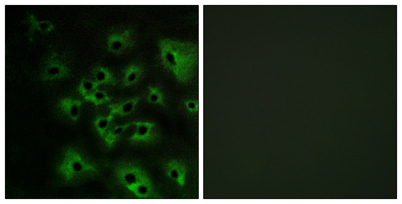
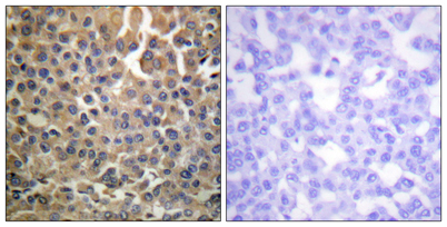
| Applications | WB;IHC;IF;ELISA |
| Other name | ERBB4; HER4; Receptor tyrosine-protein kinase erbB-4; Proto-oncogene-like protein c-ErbB-4; Tyrosine kinase-type cell surface receptor HER4; p180erbB4 |
| Size | 50μL |
| Unit price ($) | 148 |
| Human gene ID | 2066 |
| Human Swiss-Prot | Q15303 |
| Source | Rabbit |
| Isotype | IgG |
| Target | HER4/ErbB4 |
| Fields | >>MAPK signaling pathway;>>ErbB signaling pathway;>>Calcium signaling pathway;>>PI3K-Akt signaling pathway;>>Amyotrophic lateral sclerosis;>>Proteoglycans in cancer |
| Gene name | ERBB4 |
| Protein name | Receptor tyrosine-protein kinase erbB-4 |
| Human gene link | View Human Gene |
| Human Swiss link | View Human Swiss-Prot |
| Mouse gene ID | 13869 |
| Mouse gene link | View Mouse Gene |
| Mouse Swiss-Prot | Q61527 |
| Mouse Swiss link | View Mouse Swiss-Prot |
| Rat gene ID | 59323 |
| Rat gene link | View Rat Gene |
| Rat Swiss-Prot | Q62956 |
| Rat Swiss link | View Rat Swiss-Prot |
| Immunogen | The antiserum was produced against synthesized peptide derived from human HER4 around the phosphorylation site of Tyr1284. AA range:1250-1299 |
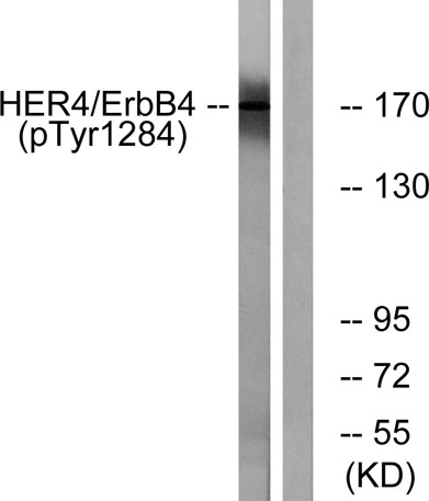
| Specificity | Phospho-ErbB-4 (Y1284) Polyclonal Antibody detects endogenous levels of ErbB-4 protein only when phosphorylated at Y1284. |
| Formulation | Liquid in PBS containing 50% glycerol, 0.5% BSA and 0.02% sodium azide. |
| Clonality | Polyclonal |
| Dilution | Western Blot: 1/500 - 1/2000. Immunohistochemistry: 1/100 - 1/300. Immunofluorescence: 1/200 - 1/1000. ELISA: 1/10000. Not yet tested in other applications. |
| Purification | The antibody was affinity-purified from rabbit antiserum by affinity-chromatography using epitope-specific immunogen. |
| Concentration | 1 mg/ml |
| Storage stability | -20°C/1 year |
| Molecular Weight (Da) | |
| Observed band (KD) | 180kD |
| Background | This gene is a member of the Tyr protein kinase family and the epidermal growth factor receptor subfamily. It encodes a single-pass type I membrane protein with multiple cysteine rich domains, a transmembrane domain, a tyrosine kinase domain, a phosphotidylinositol-3 kinase binding site and a PDZ domain binding motif. The protein binds to and is activated by neuregulins and other factors and induces a variety of cellular responses including mitogenesis and differentiation. Multiple proteolytic events allow for the release of a cytoplasmic fragment and an extracellular fragment. Mutations in this gene have been associated with cancer. Alternatively spliced variants which encode different protein isoforms have been described; however, not all variants have been fully characterized. [provided by RefSeq, Jul 2008], |
| Function | catalytic activity:ATP + a [protein]-L-tyrosine = ADP + a [protein]-L-tyrosine phosphate.,domain:The WW-binding motifs mediate interaction with WWOX.,function:Specifically binds and is activated by neuregulins, NRG-2, NRG-3, heparin-binding EGF-like growth factor, betacellulin and NTAK. Interaction with these factors induces cell differentiation. Not activated by EGF, TGF-A, and amphiregulin.,PTM:Isoform JM-A is processed but not isoform JM-B. So, they respectively represent cleavable and non-cleavable forms of the receptor.,PTM:Ligand-binding increases phosphorylation on tyrosine residues.,similarity:Belongs to the protein kinase superfamily. Tyr protein kinase family. EGF receptor subfamily.,similarity:Contains 1 protein kinase domain.,subunit:Homodimer or heterodimer with each of the other ERBB receptors (Potential). Interacts with PDZ domains of DLG2, DLG3, DLG4 and the syntrophin SN |
| Subcellular location | Cell membrane ; Single-pass type I membrane protein . In response to NRG1 treatment, the activated receptor is internalized.; [ERBB4 intracellular domain]: Nucleus . Mitochondrion . Following proteolytical processing E4ICD (E4ICD1 or E4ICD2 generated from the respective isoforms) is translocated to the nucleus. Significantly more E4ICD2 than E4ICD1 is found in the nucleus. E4ICD2 colocalizes with YAP1 in the nucleus. |
| Expression | Expressed at highest levels in brain, heart, kidney, in addition to skeletal muscle, parathyroid, cerebellum, pituitary, spleen, testis and breast. Lower levels in thymus, lung, salivary gland, and pancreas. Isoform JM-A CYT-1 and isoform JM-B CYT-1 are expressed in cerebellum, but only the isoform JM-B is expressed in the heart. |

Use collapsible tabs for more detailed information that will help customers make a purchasing decision.
Ex: Shipping and return policies, size guides, and other common questions.