 Français
 English

| ELK.No | ES5204 |
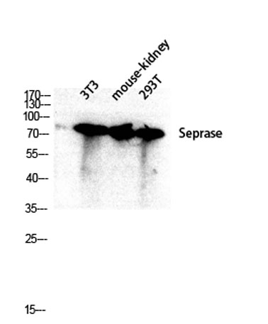
| Product name | Seprase rabbit pAb |
| Reactivity | Human;Mouse;Rat |
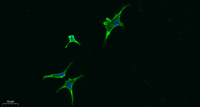
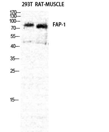
| Applications | WB;IHC;IF;ELISA |
| Other name | FAP; Seprase; 170 kDa melanoma membrane-bound gelatinase; Fibroblast activation protein alpha; Integral membrane serine protease |
| Size | 100μL |
| Unit price ($) | 248 |
| Human gene ID | 2191 |
| Human Swiss-Prot | Q12884 |
| Source | Rabbit |
| Isotype | IgG |
| Target | Seprase |
| Fields | |
| Gene name | FAP |
| Protein name | Seprase |
| Human gene link | View Human Gene |
| Human Swiss link | View Human Swiss-Prot |
| Mouse gene ID | 14089 |
| Mouse gene link | View Mouse Gene |
| Mouse Swiss-Prot | P97321 |
| Mouse Swiss link | View Mouse Swiss-Prot |
| Rat gene ID | |
| Rat gene link | |
| Rat Swiss-Prot | |
| Rat Swiss link | |
| Immunogen | The antiserum was produced against synthesized peptide derived from human FAP-1. AA range:331-380 |
| Specificity | Seprase Polyclonal Antibody detects endogenous levels of Seprase protein. |
| Formulation | Liquid in PBS containing 50% glycerol, 0.5% BSA and 0.02% sodium azide. |
| Clonality | Polyclonal |
| Dilution | Western Blot: 1/500 - 1/2000. IHC-p: 1:100-300 ELISA: 1/20000. IF 1:100-300 Not yet tested in other applications. |
| Purification | The antibody was affinity-purified from rabbit antiserum by affinity-chromatography using epitope-specific immunogen. |
| Concentration | 1 mg/ml |
| Storage stability | -20°C/1 year |
| Molecular Weight (Da) | |
| Observed band (KD) | 90kD |
| Background | The protein encoded by this gene is a homodimeric integral membrane gelatinase belonging to the serine protease family. It is selectively expressed in reactive stromal fibroblasts of epithelial cancers, granulation tissue of healing wounds, and malignant cells of bone and soft tissue sarcomas. This protein is thought to be involved in the control of fibroblast growth or epithelial-mesenchymal interactions during development, tissue repair, and epithelial carcinogenesis. Alternatively spliced transcript variants encoding different isoforms have been found for this gene. [provided by RefSeq, Apr 2014], |
| Function | catalytic activity:Degrades gelatin and heat-denatured type I and type IV collagen, but not native type I or type IV collagen. Does not cleave laminin, fibronectin, fibrin or casein.,function:May have a role in tissue remodeling during development and wound healing, and may contribute to invasiveness in malignant cancers.,induction:In fibroblasts at times and sites of tissue remodeling during development, tissue repair, and carcinogenesis.,PTM:N-glycosylated.,PTM:The N-terminus may be blocked.,similarity:Belongs to the peptidase S9B family.,subcellular location:Found in cell surface lamellipodia, invadopodia and on shed vesicles.,subunit:Homodimer, or heterodimer with DPP4. The monomer is inactive.,tissue specificity:Fibroblast specific., |
| Subcellular location | [Prolyl endopeptidase FAP]: Cell surface . Cell membrane ; Single-pass type II membrane protein . Cell projection, lamellipodium membrane ; Single-pass type II membrane protein . Cell projection, invadopodium membrane ; Single-pass type II membrane protein . Cell projection, ruffle membrane ; Single-pass type II membrane protein . Membrane ; Single-pass type II membrane protein . Localized on cell surface with lamellipodia and invadopodia membranes and on shed vesicles. Colocalized with DPP4 at invadopodia and lamellipodia membranes of migratory activated endothelial cells in collagenous matrix. Colocalized with DPP4 on endothelial cells of capillary-like microvessels but not large vessels within invasive breast ductal carcinoma. Anchored and enriched preferentially by integrin alpha-3/bet |
| Expression | Expressed in adipose tissue. Expressed in the dermal fibroblasts in the fetal skin. Expressed in the granulation tissue of healing wounds and on reactive stromal fibroblast in epithelial cancers. Expressed in activated fibroblast-like synoviocytes from inflamed synovial tissues. Expressed in activated hepatic stellate cells (HSC) and myofibroblasts from cirrhotic liver, but not detected in normal liver. Expressed in glioma cells (at protein level). Expressed in glioblastomas and glioma cells. Isoform 1 and isoform 2 are expressed in melanoma, carcinoma and fibroblast cell lines. |

Use collapsible tabs for more detailed information that will help customers make a purchasing decision.
Ex: Shipping and return policies, size guides, and other common questions.