 Français
 English
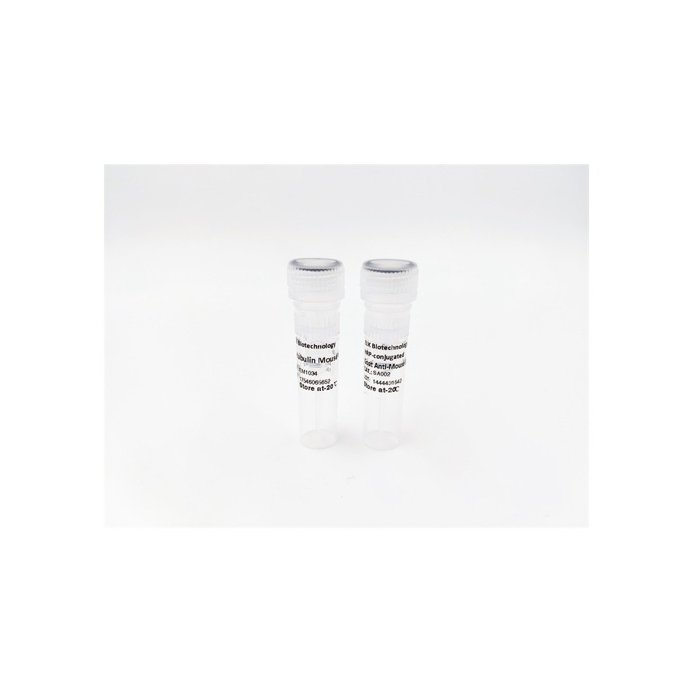

| ELK.No | ES5249 |
| Product name | FGFR-3 rabbit pAb |
| Reactivity | Human;Mouse;Rat |
| Applications | WB;IHC;IF;ELISA |
| Other name | FGFR3; JTK4; Fibroblast growth factor receptor 3; FGFR-3; CD antigen CD333 |
| Size | 50μL |
| Unit price ($) | 148 |
| Human gene ID | 2261 |
| Human Swiss-Prot | P22607 |
| Source | Rabbit |
| Isotype | IgG |
| Target | FGFR3 |
| Fields | >>EGFR tyrosine kinase inhibitor resistance;>>MAPK signaling pathway;>>Ras signaling pathway;>>Rap1 signaling pathway;>>Calcium signaling pathway;>>Endocytosis;>>PI3K-Akt signaling pathway;>>Signaling pathways regulating pluripotency of stem cells;>>Regulation of actin cytoskeleton;>>Pathways in cancer;>>MicroRNAs in cancer;>>Bladder cancer;>>Central carbon metabolism in cancer |
| Gene name | FGFR3 |
| Protein name | Fibroblast growth factor receptor 3 |
| Human gene link | View Human Gene |
| Human Swiss link | View Human Swiss-Prot |
| Mouse gene ID | |
| Mouse gene link | |
| Mouse Swiss-Prot | Q61851 |
| Mouse Swiss link | View Mouse Swiss-Prot |
| Rat gene ID | |
| Rat gene link | |
| Rat Swiss-Prot | |
| Rat Swiss link | |
| Immunogen | The antiserum was produced against synthesized peptide derived from human FGFR3. AA range:131-180 |
| Specificity | FGFR-3 Polyclonal Antibody detects endogenous levels of FGFR-3 protein. |
| Formulation | Liquid in PBS containing 50% glycerol, 0.5% BSA and 0.02% sodium azide. |
| Clonality | Polyclonal |
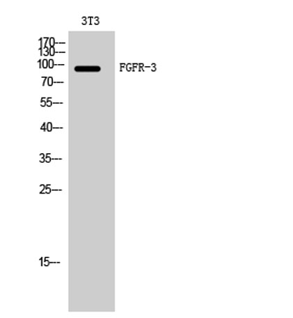
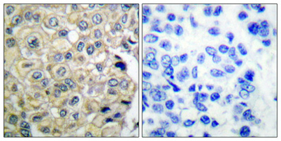
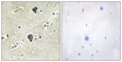
| Dilution | Western Blot: 1/500 - 1/2000. Immunohistochemistry: 1/100 - 1/300. ELISA: 1/10000. Not yet tested in other applications. |
| Purification | The antibody was affinity-purified from rabbit antiserum by affinity-chromatography using epitope-specific immunogen. |
| Concentration | 1 mg/ml |
| Storage stability | -20°C/1 year |
| Molecular Weight (Da) | |
| Observed band (KD) | 95-130kD |
| Background | This gene encodes a member of the fibroblast growth factor receptor (FGFR) family, with its amino acid sequence being highly conserved between members and among divergent species. FGFR family members differ from one another in their ligand affinities and tissue distribution. A full-length representative protein would consist of an extracellular region, composed of three immunoglobulin-like domains, a single hydrophobic membrane-spanning segment and a cytoplasmic tyrosine kinase domain. The extracellular portion of the protein interacts with fibroblast growth factors, setting in motion a cascade of downstream signals, ultimately influencing mitogenesis and differentiation. This particular family member binds acidic and basic fibroblast growth hormone and plays a role in bone development and maintenance. Mutations in this gene lead to craniosynostosis and multiple types of skeletal dys |
| Function | catalytic activity:ATP + a [protein]-L-tyrosine = ADP + a [protein]-L-tyrosine phosphate.,disease:A chromosomal aberration involving FGFR3 may be a cause of multiple myeloma (MM) [MIM:254500]. Translocation t(4;14)(p16.3;q32.3) with the IgH locus.,disease:Defects in FGFR3 are a cause of bladder cancer [MIM:109800]. Somatic mutations can constitutively activate FGFR3.,disease:Defects in FGFR3 are a cause of cervical cancer [MIM:603956].,disease:Defects in FGFR3 are a cause of hypochondroplasia (HCH) [MIM:146000]. HCH is an autosomal dominant disease and is characterized by disproportionate short stature. It resembles achondroplasia, but with a less severe phenotype.,disease:Defects in FGFR3 are a cause of keratinocytic non-epidermolytic nevus [MIM:162900]; also called pigmented moles. Epidermal nevi of the common, non-organoid and non-epidermolytic type are benign skin lesions and may var |
| Subcellular location | [Isoform 1]: Cell membrane; Single-pass type I membrane protein. Cytoplasmic vesicle. Endoplasmic reticulum. The activated receptor is rapidly internalized and degraded. Detected in intracellular vesicles after internalization of the autophosphorylated receptor.; [Isoform 2]: Cell membrane ; Single-pass type I membrane protein .; [Isoform 3]: Secreted.; [Isoform 4]: Cell membrane ; Single-pass type I membrane protein . |
| Expression | Expressed in brain, kidney and testis. Very low or no expression in spleen, heart, and muscle. In 20- to 22-week old fetuses it is expressed at high level in kidney, lung, small intestine and brain, and to a lower degree in spleen, liver, and muscle. Isoform 2 is detected in epithelial cells. Isoform 1 is not detected in epithelial cells. Isoform 1 and isoform 2 are detected in fibroblastic cells. |

Use collapsible tabs for more detailed information that will help customers make a purchasing decision.
Ex: Shipping and return policies, size guides, and other common questions.