 Français
 English
Hck (phospho Tyr410) rabbit pAb
AO-06-ES5721-100

| ELK.No | ES5721 |
| Product name | Hck (phospho Tyr410) rabbit pAb |
| Reactivity | Human;Mouse;Rat |
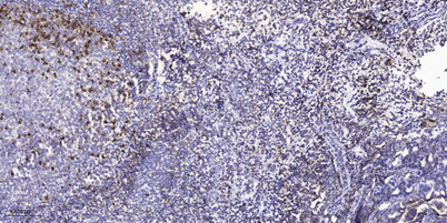
| Applications | IHC;IF;ELISA |
| Other name | HCK; Tyrosine-protein kinase HCK; Hematopoietic cell kinase; Hemopoietic cell kinase; p59-HCK/p60-HCK; p59Hck; p61Hck |
| Size | 100μL |
| Unit price ($) | 248 |
| Human gene ID | 3055 |
| Human Swiss-Prot | P08631 |
| Source | Rabbit |
| Isotype | IgG |
| Target | Hck |
| Fields | >>Chemokine signaling pathway;>>Fc gamma R-mediated phagocytosis;>>Kaposi sarcoma-associated herpesvirus infection |
| Gene name | HCK |
| Protein name | Tyrosine-protein kinase HCK |
| Human gene link | View Human Gene |
| Human Swiss link | View Human Swiss-Prot |
| Mouse gene ID | 15162 |
| Mouse gene link | View Mouse Gene |
| Mouse Swiss-Prot | P08103 |
| Mouse Swiss link | View Mouse Swiss-Prot |
| Rat gene ID | 25734 |
| Rat gene link | View Rat Gene |
| Rat Swiss-Prot | P50545 |
| Rat Swiss link | View Rat Swiss-Prot |
| Immunogen | The antiserum was produced against synthesized peptide derived from human HCK around the phosphorylation site of Tyr410. AA range:381-430 |
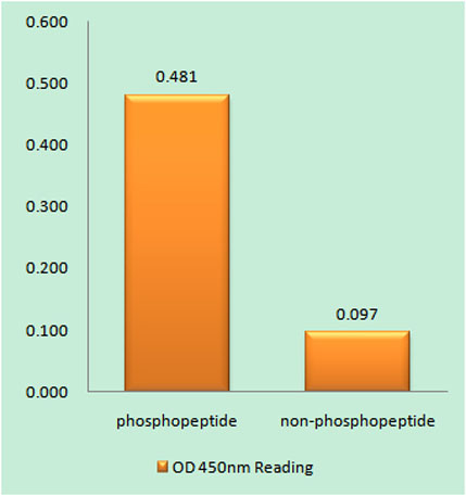
| Specificity | Phospho-Hck (Y410) Polyclonal Antibody detects endogenous levels of Hck protein only when phosphorylated at Y410. |
| Formulation | Liquid in PBS containing 50% glycerol, 0.5% BSA and 0.02% sodium azide. |
| Clonality | Polyclonal |
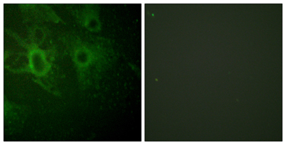
| Dilution | Immunohistochemistry: 1/100 - 1/300. Immunofluorescence: 1/200 - 1/1000. ELISA: 1/20000. Not yet tested in other applications. |
| Purification | The antibody was affinity-purified from rabbit antiserum by affinity-chromatography using epitope-specific immunogen. |
| Concentration | 1 mg/ml |
| Storage stability | -20°C/1 year |
| Molecular Weight (Da) | 60kD |
| Observed band (KD) | |
| Background | The protein encoded by this gene is a member of the Src family of tyrosine kinases. This protein is primarily hemopoietic, particularly in cells of the myeloid and B-lymphoid lineages. It may help couple the Fc receptor to the activation of the respiratory burst. In addition, it may play a role in neutrophil migration and in the degranulation of neutrophils. Multiple isoforms with different subcellular distributions are produced due to both alternative splicing and the use of alternative translation initiation codons, including a non-AUG (CUG) codon. [provided by RefSeq, Feb 2010], |
| Function | catalytic activity:ATP + a [protein]-L-tyrosine = ADP + a [protein]-L-tyrosine phosphate.,domain:The SH3 domain mediates binding to HIV-1 Nef.,function:May serve as part of a signaling pathway coupling the Fc receptor to the activation of the respiratory burst. May also contribute to neutrophil migration and may regulate the degranulation process of neutrophils.,PTM:Isoform p59-HCK contains a N-myristoyl glycine at position 3 (By similarity). Isoform p59-HCK contains a S-palmitoyl cysteine at position 3.,similarity:Belongs to the protein kinase superfamily. Tyr protein kinase family. SRC subfamily.,similarity:Contains 1 protein kinase domain.,similarity:Contains 1 SH2 domain.,similarity:Contains 1 SH3 domain.,subunit:May interact (via SH3 domain) with HIV-1 Nef and Vif. This interaction would stimulates its tyrosine-kinase activity. Interacts (via SH3 domain) with HEV ORF3 protein.,tissu |
| Subcellular location | [Isoform 1]: Lysosome. Membrane; Lipid-anchor. Cell projection, podosome membrane; Lipid-anchor. Cytoplasm, cytosol. Associated with specialized secretory lysosomes called azurophil granules. At least half of this isoform is found in the cytoplasm, some of this fraction is myristoylated.; [Isoform 2]: Cell membrane ; Lipid-anchor . Membrane, caveola ; Lipid-anchor . Cell junction, focal adhesion . Cytoplasm, cytoskeleton . Golgi apparatus . Cytoplasmic vesicle . Lysosome . Nucleus . 20% of this isoform is associated with caveolae. Localization at the cell membrane and at caveolae requires palmitoylation at Cys-3. Colocalizes with the actin cytoskeleton at focal adhesions.; Cytoplasmic vesicle, secretory vesicle. Cytoplasm, cytosol. |
| Expression | Detected in monocytes and neutrophils (at protein level). Expressed predominantly in cells of the myeloid and B-lymphoid lineages. Highly expressed in granulocytes. Detected in tonsil. |

Use collapsible tabs for more detailed information that will help customers make a purchasing decision.
Ex: Shipping and return policies, size guides, and other common questions.