 Français
 English

| ELK.No | ES6020 |
| Product name | Flk-1/Flt-4 rabbit pAb |
| Reactivity | Human;Mouse;Rat |
| Applications | IHC;IF;ELISA |
| Other name | KDR; FLK1; VEGFR2; Vascular endothelial growth factor receptor 2; VEGFR-2; Fetal liver kinase 1; FLK-1; Kinase insert domain receptor; KDR; Protein-tyrosine kinase receptor flk-1; CD antigen CD309; FLT4; VEGFR3; Vascular endothelial growth |
| Size | 100μL |
| Unit price ($) | 248 |
| Human gene ID | 3791/2324 |
| Human Swiss-Prot | P35968/P35916 |
| Source | Rabbit |
| Isotype | IgG |
| Target | VEGFR2/VEGFR3 |
| Fields | >>EGFR tyrosine kinase inhibitor resistance;>>MAPK signaling pathway;>>Ras signaling pathway;>>Rap1 signaling pathway;>>Calcium signaling pathway;>>PI3K-Akt signaling pathway;>>VEGF signaling pathway;>>Focal adhesion;>>Proteoglycans in cancer;>>Fluid shear stress and atherosclerosis |
| Gene name | KDR/FLT4 |
| Protein name | Vascular endothelial growth factor receptor 2/3 |
| Human gene link | View Human Gene |
| Human Swiss link | View Human Swiss-Prot |
| Mouse gene ID | 14257 |
| Mouse gene link | View Mouse Gene |
| Mouse Swiss-Prot | |
| Mouse Swiss link | |
| Rat gene ID | 25589/114110 |
| Rat gene link | View Rat Gene |
| Rat Swiss-Prot | O08775/Q91ZT1 |
| Rat Swiss link | View Rat Swiss-Prot |
| Immunogen | The antiserum was produced against synthesized peptide derived from human VEGFR2. AA range:1020-1069 |
| Specificity | Flk-1/Flt-4 Polyclonal Antibody detects endogenous levels of Flk-1/Flt-4 protein. |
| Formulation | Liquid in PBS containing 50% glycerol, 0.5% BSA and 0.02% sodium azide. |
| Clonality | Polyclonal |
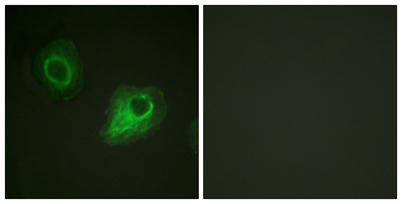
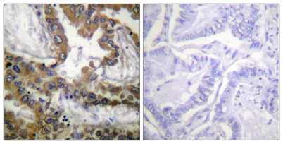
| Dilution | Immunohistochemistry: 1/100 - 1/300. Immunofluorescence: 1/200 - 1/1000. ELISA: 1/40000. Not yet tested in other applications. |
| Purification | The antibody was affinity-purified from rabbit antiserum by affinity-chromatography using epitope-specific immunogen. |
| Concentration | 1 mg/ml |
| Storage stability | -20°C/1 year |
| Molecular Weight (Da) | 152kD |
| Observed band (KD) | |
| Background | Vascular endothelial growth factor (VEGF) is a major growth factor for endothelial cells. This gene encodes one of the two receptors of the VEGF. This receptor, known as kinase insert domain receptor, is a type III receptor tyrosine kinase. It functions as the main mediator of VEGF-induced endothelial proliferation, survival, migration, tubular morphogenesis and sprouting. The signalling and trafficking of this receptor are regulated by multiple factors, including Rab GTPase, P2Y purine nucleotide receptor, integrin alphaVbeta3, T-cell protein tyrosine phosphatase, etc.. Mutations of this gene are implicated in infantile capillary hemangiomas. [provided by RefSeq, May 2009], |
| Function | catalytic activity:ATP + a [protein]-L-tyrosine = ADP + a [protein]-L-tyrosine phosphate.,function:Receptor for VEGF or VEGFC. Has a tyrosine-protein kinase activity. The VEGF-kinase ligand/receptor signaling system plays a key role in vascular development and regulation of vascular permeability. In case of HIV-1 infection, the interaction with extracellular viral Tat protein seems to enhance angiogenesis in Kaposi's sarcoma lesions.,similarity:Belongs to the protein kinase superfamily. Tyr protein kinase family.,similarity:Belongs to the protein kinase superfamily. Tyr protein kinase family. CSF-1/PDGF receptor subfamily.,similarity:Contains 1 protein kinase domain.,similarity:Contains 7 Ig-like C2-type (immunoglobulin-like) domains.,subunit:Interacts with MYOF (By similarity). Interacts with SHB; upon VEGF activation. Interacts with HIV-1 Tat., |
| Subcellular location | Cell junction . Endoplasmic reticulum . Cell membrane . Localized with RAP1A at cell-cell junctions (By similarity). Colocalizes with ERN1 and XBP1 in the endoplasmic reticulum in endothelial cells in a vascular endothelial growth factor (VEGF)-dependent manner (PubMed:23529610). .; [Isoform 1]: Cell membrane; Single-pass type I membrane protein. Cytoplasm. Nucleus. Cytoplasmic vesicle. Early endosome. Detected on caveolae-enriched lipid rafts at the cell surface. Is recycled from the plasma membrane to endosomes and back again. Phosphorylation triggered by VEGFA binding promotes internalization and subsequent degradation. VEGFA binding triggers internalization and translocation to the nucleus.; [Isoform 2]: Secreted .; [Isoform 3]: Secreted. |
| Expression | Detected in cornea (at protein level). Widely expressed. |

Use collapsible tabs for more detailed information that will help customers make a purchasing decision.
Ex: Shipping and return policies, size guides, and other common questions.