 Français
 English

| ELK.No | ES6075 |
| Product name | Laminin α-3 rabbit pAb |
| Reactivity | Human;Mouse;Rat |
| Applications | WB;IHC;IF;ELISA |
| Other name | LAMA3; LAMNA; Laminin subunit alpha-3; Epiligrin 170 kDa subunit; E170; Epiligrin subunit alpha; Kalinin subunit alpha; Laminin-5 subunit alpha; Laminin-6 subunit alpha; Laminin-7 subunit alpha; Nicein subunit alpha |
| Size | 50μL |
| Unit price ($) | 148 |
| Human gene ID | 3909 |
| Human Swiss-Prot | Q16787 |
| Source | Rabbit |
| Isotype | IgG |
| Target | Laminin α-3 |
| Fields | >>PI3K-Akt signaling pathway;>>Focal adhesion;>>ECM-receptor interaction;>>Toxoplasmosis;>>Amoebiasis;>>Human papillomavirus infection;>>Pathways in cancer;>>Small cell lung cancer |
| Gene name | LAMA3 |
| Protein name | Laminin subunit alpha-3 |
| Human gene link | View Human Gene |
| Human Swiss link | View Human Swiss-Prot |
| Mouse gene ID | |
| Mouse gene link | |
| Mouse Swiss-Prot | Q61789 |
| Mouse Swiss link | View Mouse Swiss-Prot |
| Rat gene ID | |
| Rat gene link | |
| Rat Swiss-Prot | |
| Rat Swiss link | |
| Immunogen | The antiserum was produced against synthesized peptide derived from human LAMA3. AA range:2571-2620 |
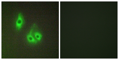
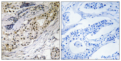
| Specificity | Laminin α-3 Polyclonal Antibody detects endogenous levels of Laminin α-3 protein. |
| Formulation | Liquid in PBS containing 50% glycerol, 0.5% BSA and 0.02% sodium azide. |
| Clonality | Polyclonal |
| Dilution | Western Blot: 1/500 - 1/2000. Immunohistochemistry: 1/100 - 1/300. Immunofluorescence: 1/200 - 1/1000. ELISA: 1/40000. Not yet tested in other applications. |
| Purification | The antibody was affinity-purified from rabbit antiserum by affinity-chromatography using epitope-specific immunogen. |
| Concentration | 1 mg/ml |
| Storage stability | -20°C/1 year |
| Molecular Weight (Da) | 343kD |
| Observed band (KD) | 120 60kD |
| Background | The protein encoded by this gene belongs to the laminin family of secreted molecules. Laminins are heterotrimeric molecules that consist of alpha, beta, and gamma subunits that assemble through a coiled-coil domain. Laminins are essential for formation and function of the basement membrane and have additional functions in regulating cell migration and mechanical signal transduction. This gene encodes an alpha subunit and is responsive to several epithelial-mesenchymal regulators including keratinocyte growth factor, epidermal growth factor and insulin-like growth factor. Mutations in this gene have been identified as the cause of Herlitz type junctional epidermolysis bullosa and laryngoonychocutaneous syndrome. Alternative splicing and alternative promoter usage result in multiple transcript variants. [provided by RefSeq, Dec 2014], |
| Function | disease:Defects in LAMA3 are a cause of epidermolysis bullosa junctional Herlitz type (H-JEB) [MIM:226700]; also known as junctional epidermolysis bullosa Herlitz-Pearson type. JEB defines a group of blistering skin diseases characterized by tissue separation which occurs within the dermo-epidermal basement membrane. H-JEB is a severe, infantile and lethal form. Death occurs usually within the first six months of life. Occasionally, children survive to teens. H-JEB is marked by bullous lesions at birth and extensive denudation of skin and mucous membranes that may be hemorrhagic.,disease:Defects in LAMA3 are the cause of laryngoonychocutaneous syndrome (LOCS) [MIM:245660]. LOCS is an autosomal recessive epithelial disorder confined to the Punjabi Muslim population. The condition is characterized by cutaneous erosions, nail dystrophy and exuberant vascular granulation tissue in certain ep |
| Subcellular location | Secreted, extracellular space, extracellular matrix, basement membrane. Major component. |
| Expression | Skin; respiratory, urinary, and digestive epithelia and in other specialized tissues with prominent secretory or protective functions. Epithelial basement membrane, and epithelial cell tongue that migrates into a wound bed. A differential and focal expression of the subunit alpha-3 is observed in the CNS. |

Use collapsible tabs for more detailed information that will help customers make a purchasing decision.
Ex: Shipping and return policies, size guides, and other common questions.