 Français
 English
Mucin 1 (phospho Tyr1229) rabbit pAb
AO-06-ES6288-50

| ELK.No | ES6288 |
| Product name | Mucin 1 (phospho Tyr1229) rabbit pAb |
| Reactivity | Human;Rat;Mouse; |
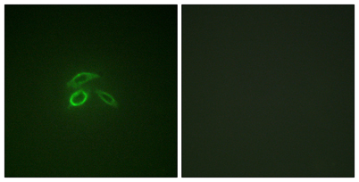
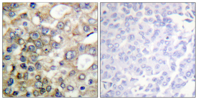
| Applications | WB;IHC;IF;ELISA |
| Other name | MUC1; PUM; Mucin-1; MUC-1; Breast carcinoma-associated antigen DF3; Carcinoma-associated mucin; Episialin; H23AG; Krebs von den Lungen-6; KL-6; PEMT; Peanut-reactive urinary mucin; PUM; Polymorphic epithelial mucin; PEM; Tumor-associated ep |
| Size | 50μL |
| Unit price ($) | 148 |
| Human gene ID | 4582 |
| Human Swiss-Prot | P15941 |
| Source | Rabbit |
| Isotype | IgG |
| Target | Mucin 1 |
| Fields | |
| Gene name | MUC1 |
| Protein name | Mucin-1 (MUC-1) (Breast carcinoma-associated antigen DF3) (Carcinoma-associated mucin) (Episialin) (H23AG) (Krebs von den Lungen-6) (KL-6) (PEMT) (Peanut-reactive urinary mucin) (PUM) (Polymorphic epi |
| Human gene link | View Human Gene |
| Human Swiss link | View Human Swiss-Prot |
| Mouse gene ID | |
| Mouse gene link | |
| Mouse Swiss-Prot | Q02496 |
| Mouse Swiss link | View Mouse Swiss-Prot |
| Rat gene ID | |
| Rat gene link | |
| Rat Swiss-Prot | |
| Rat Swiss link | |
| Immunogen | The antiserum was produced against synthesized peptide derived from human CD227/MUC1 around the phosphorylation site of Tyr1229. AA range:1201-1250 |
| Specificity | Phospho-Mucin 1 (Y1229) Polyclonal Antibody detects endogenous levels of Mucin 1 protein only when phosphorylated at Y1229. |
| Formulation | Liquid in PBS containing 50% glycerol, 0.5% BSA and 0.02% sodium azide. |
| Clonality | Polyclonal |
| Dilution | Western Blot: 1/500 - 1/2000. Immunohistochemistry: 1/100 - 1/300. Immunofluorescence: 1/200 - 1/1000. ELISA: 1/10000. Not yet tested in other applications. |
| Purification | The antibody was affinity-purified from rabbit antiserum by affinity-chromatography using epitope-specific immunogen. |
| Concentration | 1 mg/ml |
| Storage stability | -20°C/1 year |
| Molecular Weight (Da) | |
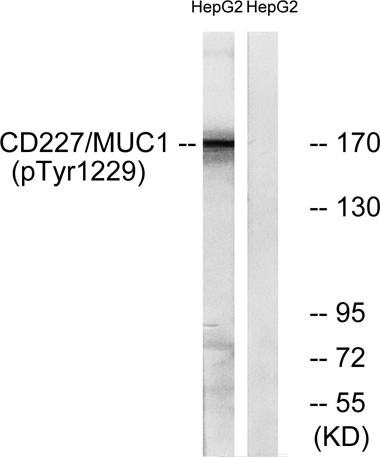
| Observed band (KD) | 170kD |
| Background | This gene encodes a membrane-bound protein that is a member of the mucin family. Mucins are O-glycosylated proteins that play an essential role in forming protective mucous barriers on epithelial surfaces. These proteins also play a role in intracellular signaling. This protein is expressed on the apical surface of epithelial cells that line the mucosal surfaces of many different tissues including lung, breast stomach and pancreas. This protein is proteolytically cleaved into alpha and beta subunits that form a heterodimeric complex. The N-terminal alpha subunit functions in cell-adhesion and the C-terminal beta subunit is involved in cell signaling. Overexpression, aberrant intracellular localization, and changes in glycosylation of this protein have been associated with carcinomas. This gene is known to contain a highly polymorphic variable number tandem repeats (VNTR) domain. Alternate sp |
| Function | alternative products:Additional isoforms seem to exist,caution:O-glycosylation sites are annotated in first sequence repeat only. Residues at similar position are probably glycosylated in all repeats. Experimental sites were determined in a synthetic peptide glycosylated in vitro (PubMed:7744025, PubMed:9597769).,caution:The N-terminal sequence has been shown (PubMed:11341784) to begin at position 24 or 28.,developmental stage:During fetal development, expressed at low levels in the colonic epithelium from 13 weeks of gestation.,function:The alpha subunit has cell adhesive properties. Can act both as an adhesion and an anti-adhesion protein. May provide a protective layer on epithelial cells against bacterial and enzyme attack.,function:The beta subunit contains a C-terminal domain which is involved in cell signaling, through phosphorylations and protein-protein interactions. Modulates s |
| Subcellular location | Apical cell membrane ; Single-pass type I membrane protein . Exclusively located in the apical domain of the plasma membrane of highly polarized epithelial cells. After endocytosis, internalized and recycled to the cell membrane. Located to microvilli and to the tips of long filopodial protusions.; [Isoform 5]: Secreted.; [Isoform Y]: Secreted.; [Isoform 9]: Secreted.; [Mucin-1 subunit beta]: Cell membrane. Cytoplasm. Nucleus. On EGF and PDGFRB stimulation, transported to the nucleus through interaction with CTNNB1, a process which is stimulated by phosphorylation. On HRG stimulation, colocalizes with JUP/gamma-catenin at the nucleus. |
| Expression | Expressed on the apical surface of epithelial cells, especially of airway passages, breast and uterus. Also expressed in activated and unactivated T-cells. Overexpressed in epithelial tumors, such as breast or ovarian cancer and also in non-epithelial tumor cells. Isoform Y is expressed in tumor cells only. |

Use collapsible tabs for more detailed information that will help customers make a purchasing decision.
Ex: Shipping and return policies, size guides, and other common questions.