 Français
 English

| ELK.No | ES6317 |
| Product name | MYLK rabbit pAb |
| Reactivity | Human;Mouse;Rat |
| Applications | WB;ELISA;IHC |
| Other name | MYLK; MLCK; MLCK1; MYLK1; Myosin light chain kinase; smooth muscle; MLCK; smMLCK; Kinase-related protein; KRP; Telokin |
| Size | 50μL |
| Unit price ($) | 148 |
| Human gene ID | 4638 |
| Human Swiss-Prot | Q15746 |
| Source | Rabbit |
| Isotype | IgG |
| Target | MYLK |
| Fields | >>Calcium signaling pathway;>>cGMP-PKG signaling pathway;>>Vascular smooth muscle contraction;>>Apelin signaling pathway;>>Focal adhesion;>>Platelet activation;>>Regulation of actin cytoskeleton;>>Oxytocin signaling pathway;>>Gastric acid secretion |
| Gene name | MYLK |
| Protein name | Myosin light chain kinase smooth muscle |
| Human gene link | View Human Gene |
| Human Swiss link | View Human Swiss-Prot |
| Mouse gene ID | |
| Mouse gene link | |
| Mouse Swiss-Prot | Q6PDN3 |
| Mouse Swiss link | View Mouse Swiss-Prot |
| Rat gene ID | |
| Rat gene link | |
| Rat Swiss-Prot | |
| Rat Swiss link | |
| Immunogen | The antiserum was produced against synthesized peptide derived from human MYLK. AA range:1701-1750 |
| Specificity | MYLK Polyclonal Antibody detects endogenous levels of MYLK protein. |
| Formulation | Liquid in PBS containing 50% glycerol, 0.5% BSA and 0.02% sodium azide. |
| Clonality | Polyclonal |
| Dilution | WB 1:500-2000;IHC-p 1:50-300; ELISA 2000-20000 |
| Purification | The antibody was affinity-purified from rabbit antiserum by affinity-chromatography using epitope-specific immunogen. |
| Concentration | 1 mg/ml |
| Storage stability | -20°C/1 year |
| Molecular Weight (Da) | |
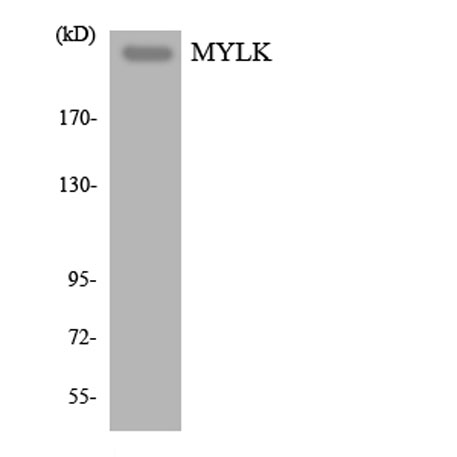
| Observed band (KD) | 200kD |
| Background | myosin light chain kinase(MYLK) Homo sapiens This gene, a muscle member of the immunoglobulin gene superfamily, encodes myosin light chain kinase which is a calcium/calmodulin dependent enzyme. This kinase phosphorylates myosin regulatory light chains to facilitate myosin interaction with actin filaments to produce contractile activity. This gene encodes both smooth muscle and nonmuscle isoforms. In addition, using a separate promoter in an intron in the 3' region, it encodes telokin, a small protein identical in sequence to the C-terminus of myosin light chain kinase, that is independently expressed in smooth muscle and functions to stabilize unphosphorylated myosin filaments. A pseudogene is located on the p arm of chromosome 3. Four transcript variants that produce four isoforms of the calcium/calmodulin dependent enzyme have been identified as well as two transcripts that produce two isoforms of telokin. Additional variants have been |
| Function | alternative products:Additional isoforms seem to exist,catalytic activity:ATP + [myosin light-chain] = ADP + [myosin light-chain] phosphate.,cofactor:Calcium.,cofactor:Magnesium.,enzyme regulation:Isoform 1 is activated by phosphorylation on Tyr-464 and Tyr-471. Isoforms which lack these tyrosine residues are not regulated in this way. All catalytically active isoforms require binding to calcium and calmodulin for activation.,function:Calcium/calmodulin-dependent enzyme implicated in smooth muscle contraction via phosphorylation of myosin light chains (MLC). Implicated in the regulation of endothelial as well as vascular permeability. In the nervous system it has been shown to control the growth initiation of astrocytic processes in culture and to participate in transmitter release at synapses formed between cultured sympathetic ganglion cells. Critical participant in signaling sequences |
| Subcellular location | Cytoplasm . Cell projection, lamellipodium . Cleavage furrow . Cytoplasm, cytoskeleton, stress fiber . Localized to stress fibers during interphase and to the cleavage furrow during mitosis. . |
| Expression | Smooth muscle and non-muscle isozymes are expressed in a wide variety of adult and fetal tissues and in cultured endothelium with qualitative expression appearing to be neither tissue- nor development-specific. Non-muscle isoform 2 is the dominant splice variant expressed in various tissues. Telokin has been found in a wide variety of adult and fetal tissues. Accumulates in well differentiated enterocytes of the intestinal epithelium in response to tumor necrosis factor (TNF). |

Use collapsible tabs for more detailed information that will help customers make a purchasing decision.
Ex: Shipping and return policies, size guides, and other common questions.