 Français
 English
NFATc3 (phospho Ser165) rabbit pAb
AO-06-ES6368-50

| ELK.No | ES6368 |
| Product name | NFATc3 (phospho Ser165) rabbit pAb |
| Reactivity | Human;Mouse |
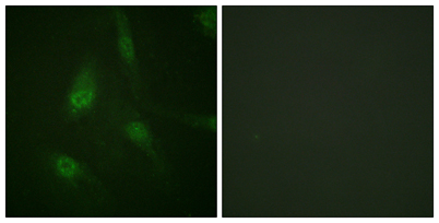
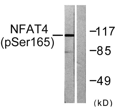
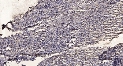
| Applications | WB;IHC;IF;ELISA |
| Other name | NFATC3; NFAT4; Nuclear factor of activated T-cells; cytoplasmic 3; NF-ATc3; NFATc3; NFATx; T-cell transcription factor NFAT4; NF-AT4 |
| Size | 50μL |
| Unit price ($) | 148 |
| Human gene ID | 4775 |
| Human Swiss-Prot | Q12968 |
| Source | Rabbit |
| Isotype | IgG |
| Target | NFAT4 |
| Fields | >>MAPK signaling pathway;>>cGMP-PKG signaling pathway;>>Cellular senescence;>>Wnt signaling pathway;>>Axon guidance;>>C-type lectin receptor signaling pathway;>>Th1 and Th2 cell differentiation;>>Th17 cell differentiation;>>T cell receptor signaling pathway;>>B cell receptor signaling pathway;>>Oxytocin signaling pathway;>>Yersinia infection;>>Hepatitis B;>>Human cytomegalovirus infection;>>Human T-cell leukemia virus 1 infection;>>Kaposi sarcoma-associated herpesvirus infection;>>Human immunodeficiency virus 1 infection;>>PD-L1 expression and PD-1 checkpoint pathway in cancer;>>Lipid and atherosclerosis |
| Gene name | NFATC3 |
| Protein name | Nuclear factor of activated T-cells cytoplasmic 3 |
| Human gene link | View Human Gene |
| Human Swiss link | View Human Swiss-Prot |
| Mouse gene ID | |
| Mouse gene link | |
| Mouse Swiss-Prot | P97305 |
| Mouse Swiss link | View Mouse Swiss-Prot |
| Rat gene ID | |
| Rat gene link | |
| Rat Swiss-Prot | |
| Rat Swiss link | |
| Immunogen | The antiserum was produced against synthesized peptide derived from human NFAT4 around the phosphorylation site of Ser165. AA range:131-180 |
| Specificity | Phospho-NFATc3 (S165) Polyclonal Antibody detects endogenous levels of NFATc3 protein only when phosphorylated at S165. |
| Formulation | Liquid in PBS containing 50% glycerol, 0.5% BSA and 0.02% sodium azide. |
| Clonality | Polyclonal |
| Dilution | Western Blot: 1/500 - 1/2000. Immunohistochemistry: 1/100 - 1/300. Immunofluorescence: 1/200 - 1/1000. ELISA: 1/5000. Not yet tested in other applications. |
| Purification | The antibody was affinity-purified from rabbit antiserum by affinity-chromatography using epitope-specific immunogen. |
| Concentration | 1 mg/ml |
| Storage stability | -20°C/1 year |
| Molecular Weight (Da) | |
| Observed band (KD) | 115kD |
| Background | The product of this gene is a member of the nuclear factors of activated T cells DNA-binding transcription complex. This complex consists of at least two components: a preexisting cytosolic component that translocates to the nucleus upon T cell receptor (TCR) stimulation and an inducible nuclear component. Other members of this family participate to form this complex also. The product of this gene plays a role in the regulation of gene expression in T cells and immature thymocytes. Several transcript variants encoding distinct isoforms have been identified for this gene. [provided by RefSeq, Nov 2010], |
| Function | domain:Rel Similarity Domain (RSD) allows DNA-binding and cooperative interactions with AP1 factors.,function:Plays a role in the inducible expression of cytokine genes in T-cells, especially in the induction of the IL-2.,PTM:Phosphorylated by NFATC-kinase; dephosphorylated by calcineurin.,similarity:Contains 1 RHD (Rel-like) domain.,subcellular location:Cytoplasmic for the phosphorylated form and nuclear after activation that is controlled by calcineurin-mediated dephosphorylation. Rapid nuclear exit of NFATC is thought to be one mechanism by which cells distinguish between sustained and transient calcium signals. The subcellular localization of NFATC plays a key role in the regulation of gene transcription.,subunit:Member of the multicomponent NFATC transcription complex that consists of at least two components, a pre-existing cytoplasmic component NFATC2 and an inducible nuclear compo |
| Subcellular location | Cytoplasm . Nucleus . Cytoplasmic for the phosphorylated form and nuclear after activation that is controlled by calcineurin-mediated dephosphorylation. Rapid nuclear exit of NFATC is thought to be one mechanism by which cells distinguish between sustained and transient calcium signals. The subcellular localization of NFATC plays a key role in the regulation of gene transcription. |
| Expression | Isoform 1 is predominantly expressed in thymus and is also found in peripheral blood leukocytes and kidney. Isoform 2 is predominantly expressed in skeletal muscle and is also found in thymus, kidney, testis, spleen, prostate, ovary, small intestine, heart, placenta and pancreas. Isoform 3 is expressed in thymus and kidney. Isoform 4 is expressed in thymus and skeletal muscle. |

Use collapsible tabs for more detailed information that will help customers make a purchasing decision.
Ex: Shipping and return policies, size guides, and other common questions.