 Français
 English
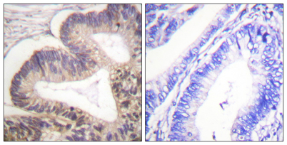
PAKγ (phospho Ser20) rabbit pAb
AO-06-ES6469-100

| ELK.No | ES6469 |
| Product name | PAKγ (phospho Ser20) rabbit pAb |
| Reactivity | Human;Mouse;Rat |
| Applications | WB;IHC;IF;ELISA |
| Other name | PAK2; Serine/threonine-protein kinase PAK 2; Gamma-PAK; PAK65; S6/H4 kinase; p21-activated kinase 2; PAK-2; p58 |
| Size | 100μL |
| Unit price ($) | 248 |
| Human gene ID | 5062 |
| Human Swiss-Prot | Q13177 |
| Source | Rabbit |
| Isotype | IgG |
| Target | PAK2 |
| Fields | >>MAPK signaling pathway;>>ErbB signaling pathway;>>Ras signaling pathway;>>Axon guidance;>>Focal adhesion;>>T cell receptor signaling pathway;>>Regulation of actin cytoskeleton;>>Pathogenic Escherichia coli infection;>>Human immunodeficiency virus 1 infection;>>Renal cell carcinoma |
| Gene name | PAK2 |
| Protein name | Serine/threonine-protein kinase PAK 2 |
| Human gene link | View Human Gene |
| Human Swiss link | View Human Swiss-Prot |
| Mouse gene ID | 224105 |
| Mouse gene link | View Mouse Gene |
| Mouse Swiss-Prot | Q8CIN4 |
| Mouse Swiss link | View Mouse Swiss-Prot |
| Rat gene ID | 1.00911e+008 |
| Rat gene link | View Rat Gene |
| Rat Swiss-Prot | Q64303 |
| Rat Swiss link | View Rat Swiss-Prot |
| Immunogen | The antiserum was produced against synthesized peptide derived from human PAK2 around the phosphorylation site of Ser20. AA range:5-54 |
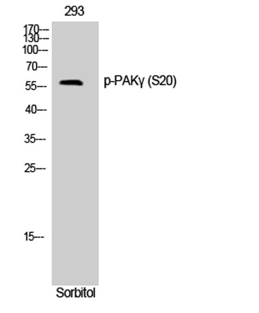
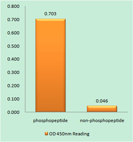
| Specificity | Phospho-PAKγ (S20) Polyclonal Antibody detects endogenous levels of PAKγ protein only when phosphorylated at S20. |
| Formulation | Liquid in PBS containing 50% glycerol, 0.5% BSA and 0.02% sodium azide. |
| Clonality | Polyclonal |
| Dilution | Western Blot: 1/500 - 1/2000. Immunohistochemistry: 1/100 - 1/300. ELISA: 1/40000. Not yet tested in other applications. |
| Purification | The antibody was affinity-purified from rabbit antiserum by affinity-chromatography using epitope-specific immunogen. |
| Concentration | 1 mg/ml |
| Storage stability | -20°C/1 year |
| Molecular Weight (Da) | |
| Observed band (KD) | 62kD |
| Background | The p21 activated kinases (PAK) are critical effectors that link Rho GTPases to cytoskeleton reorganization and nuclear signaling. The PAK proteins are a family of serine/threonine kinases that serve as targets for the small GTP binding proteins, CDC42 and RAC1, and have been implicated in a wide range of biological activities. The protein encoded by this gene is activated by proteolytic cleavage during caspase-mediated apoptosis, and may play a role in regulating the apoptotic events in the dying cell. [provided by RefSeq, Jul 2008], |
| Function | catalytic activity:ATP + a protein = ADP + a phosphoprotein.,enzyme regulation:Activated by binding small G proteins. Binding of GTP-bound CDC42 or RAC1 to the autoregulatory region releases monomers from the autoinhibited dimer, enables phosphorylation of Thr-402 and allows the kinase domain to adopt an active structure (By similarity). Following caspase cleavage, autophosphorylted PAK-2p34 is constitutively active.,function:The activated kinase acts on a variety of targets. Phosphorylates ribosomal protein S6, histone H4 and myelin basic protein. Full length PAK 2 stimulates cell survival and cell growth. The process is, at least in part, mediated by phosphorylation and inhibition of pro-apoptotic BAD. Caspase-activated PAK-2p34 is involved in cell death response, probably involving the JNK signaling pathway. Cleaved PAK-2p34 seems to have a higher activity than the CDC42-activated for |
| Subcellular location | [Serine/threonine-protein kinase PAK 2]: Cytoplasm. MYO18A mediates the cellular distribution of the PAK2-ARHGEF7-GIT1 complex to the inner surface of the cell membrane.; [PAK-2p34]: Nucleus. Cytoplasm, perinuclear region. Membrane; Lipid-anchor. Interaction with ARHGAP10 probably changes PAK-2p34 location to cytoplasmic perinuclear region. Myristoylation changes PAK-2p34 location to the membrane. |
| Expression | Ubiquitously expressed. Higher levels seen in skeletal muscle, ovary, thymus and spleen. |

Use collapsible tabs for more detailed information that will help customers make a purchasing decision.
Ex: Shipping and return policies, size guides, and other common questions.