 Français
 English
LEF-1 (phospho Ser42) rabbit pAb
AO-06-ES6501-100

| ELK.No | ES6501 |
| Product name | LEF-1 (phospho Ser42) rabbit pAb |
| Reactivity | Human;Mouse;Rat |
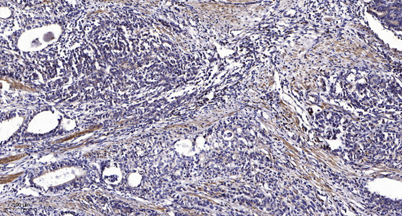
| Applications | WB;IHC |
| Other name | LEF1; Lymphoid enhancer-binding factor 1; LEF-1; T cell-specific transcription factor 1-alpha; TCF1-alpha |
| Size | 100μL |
| Unit price ($) | 248 |
| Human gene ID | 51176 |
| Human Swiss-Prot | Q9UJU2 |
| Source | Rabbit |
| Isotype | IgG |
| Target | LEF-1 |
| Fields | >>Wnt signaling pathway;>>Hippo signaling pathway;>>Adherens junction;>>Melanogenesis;>>Cushing syndrome;>>Alcoholic liver disease;>>Salmonella infection;>>Kaposi sarcoma-associated herpesvirus infection;>>Pathways in cancer;>>Colorectal cancer;>>Endometrial cancer;>>Prostate cancer;>>Thyroid cancer;>>Basal cell carcinoma;>>Acute myeloid leukemia;>>Breast cancer;>>Hepatocellular carcinoma;>>Gastric cancer;>>Arrhythmogenic right ventricular cardiomyopathy |
| Gene name | LEF1 |
| Protein name | Lymphoid enhancer-binding factor 1 |
| Human gene link | View Human Gene |
| Human Swiss link | View Human Swiss-Prot |
| Mouse gene ID | 16842 |
| Mouse gene link | View Mouse Gene |
| Mouse Swiss-Prot | P27782 |
| Mouse Swiss link | View Mouse Swiss-Prot |
| Rat gene ID | 161452 |
| Rat gene link | View Rat Gene |
| Rat Swiss-Prot | Q9QXN1 |
| Rat Swiss link | View Rat Swiss-Prot |
| Immunogen | The antiserum was produced against synthesized peptide derived from human LEF-1 around the phosphorylation site of Ser42. AA range:8-57 |
| Specificity | Phospho-LEF-1 (S42) Polyclonal Antibody detects endogenous levels of LEF-1 protein only when phosphorylated at S42. |
| Formulation | Liquid in PBS containing 50% glycerol, 0.5% BSA and 0.02% sodium azide. |
| Clonality | Polyclonal |
| Dilution | WB 1:500-2000;IHC-p 1:50-300 |
| Purification | The antibody was affinity-purified from rabbit antiserum by affinity-chromatography using epitope-specific immunogen. |
| Concentration | 1 mg/ml |
| Storage stability | -20°C/1 year |
| Molecular Weight (Da) | |
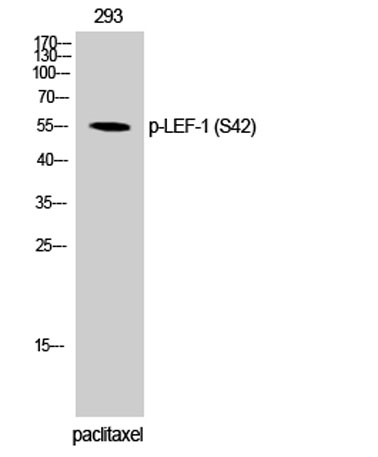
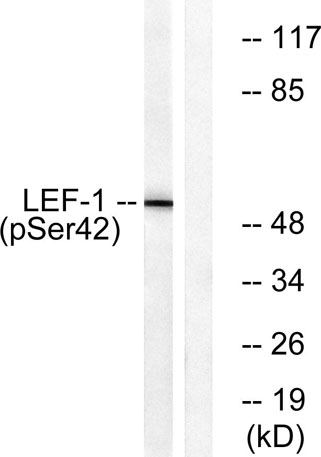
| Observed band (KD) | 55kD |
| Background | This gene encodes a transcription factor belonging to a family of proteins that share homology with the high mobility group protein-1. The protein encoded by this gene can bind to a functionally important site in the T-cell receptor-alpha enhancer, thereby conferring maximal enhancer activity. This transcription factor is involved in the Wnt signaling pathway, and it may function in hair cell differentiation and follicle morphogenesis. Mutations in this gene have been found in somatic sebaceous tumors. This gene has also been linked to other cancers, including androgen-independent prostate cancer. Alternative splicing results in multiple transcript variants. [provided by RefSeq, Oct 2009], |
| Function | alternative products:Additional isoforms seem to exist,domain:Proline-rich and acidic regions are implicated in the activation functions of RNA polymerase II transcription factors.,function:Participates in the Wnt signaling pathway. Activates transcription of target genes in the presence of CTNNB1 and EP300. May play a role in hair cell differentiation and follicle morphogenesis. TLE1, TLE2, TLE3 and TLE4 repress transactivation mediated by LEF1 and CTNNB1. Regulates T-cell receptor alpha enhancer function. Binds DNA in a sequence-specific manner. PIAG antagonizes both Wnt-dependent and Wnt-independent activation by LEF1 (By similarity). Isoform 3 lacks the CTNNB1 interaction domain and may be an antagonist for Wnt signaling.,similarity:Belongs to the TCF/LEF family.,similarity:Contains 1 HMG box DNA-binding domain.,subcellular location:Found in nuclear bodies upon PIASG binding.,subunit |
| Subcellular location | Nucleus . Found in nuclear bodies upon PIASG binding. . |
| Expression | Detected in thymus. Not detected in normal colon, but highly expressed in colon cancer biopsies and colon cancer cell lines. Expressed in several pancreatic tumors and weakly expressed in normal pancreatic tissue. Isoforms 1 and 5 are detected in several pancreatic cell lines. |

Use collapsible tabs for more detailed information that will help customers make a purchasing decision.
Ex: Shipping and return policies, size guides, and other common questions.