 Français
 English

| ELK.No | ES6620 |
| Product name | Plk rabbit pAb |
| Reactivity | Human;Mouse;Rat |
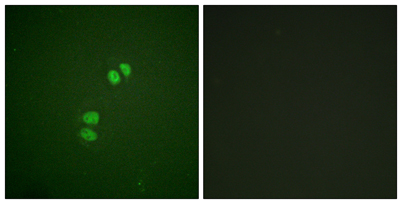
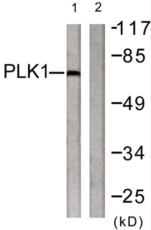
| Applications | WB;IHC;IF;ELISA |
| Other name | PLK1; PLK; Serine/threonine-protein kinase PLK1; Polo-like kinase 1; PLK-1; Serine/threonine-protein kinase 13; STPK13 |
| Size | 50μL |
| Unit price ($) | 148 |
| Human gene ID | 5347 |
| Human Swiss-Prot | P53350 |
| Source | Rabbit |
| Isotype | IgG |
| Target | PLK1 |
| Fields | >>FoxO signaling pathway;>>Cell cycle;>>Oocyte meiosis;>>Progesterone-mediated oocyte maturation |
| Gene name | PLK1 |
| Protein name | Serine/threonine-protein kinase PLK1 |
| Human gene link | View Human Gene |
| Human Swiss link | View Human Swiss-Prot |
| Mouse gene ID | 18817 |
| Mouse gene link | View Mouse Gene |
| Mouse Swiss-Prot | Q07832 |
| Mouse Swiss link | View Mouse Swiss-Prot |
| Rat gene ID | 25515 |
| Rat gene link | View Rat Gene |
| Rat Swiss-Prot | Q62673 |
| Rat Swiss link | View Rat Swiss-Prot |
| Immunogen | The antiserum was produced against synthesized peptide derived from human PLK1. AA range:176-225 |
| Specificity | Plk Polyclonal Antibody detects endogenous levels of Plk protein. |
| Formulation | Liquid in PBS containing 50% glycerol, 0.5% BSA and 0.02% sodium azide. |
| Clonality | Polyclonal |
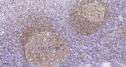
| Dilution | Western Blot: 1/500 - 1/2000. Immunohistochemistry: 1/100 - 1/300. Immunofluorescence: 1/200 - 1/1000. ELISA: 1/20000. Not yet tested in other applications. |
| Purification | The antibody was affinity-purified from rabbit antiserum by affinity-chromatography using epitope-specific immunogen. |
| Concentration | 1 mg/ml |
| Storage stability | -20°C/1 year |
| Molecular Weight (Da) | |
| Observed band (KD) | 70kD |
| Background | The Ser/Thr protein kinase encoded by this gene belongs to the CDC5/Polo subfamily. It is highly expressed during mitosis and elevated levels are found in many different types of cancer. Depletion of this protein in cancer cells dramatically inhibited cell proliferation and induced apoptosis; hence, it is a target for cancer therapy. [provided by RefSeq, Sep 2015], |
| Function | catalytic activity:ATP + a protein = ADP + a phosphoprotein.,developmental stage:Accumulates to a maximum during the G2 and M phases, declines to a nearly undetectable level following mitosis and throughout G1 phase, and then begins to accumulate again during S phase.,enzyme regulation:Activated by serine and threonine phosphorylation.,function:Serine/threonine-protein kinase that performs several important functions throughout M phase of the cell cycle, including the regulation of centrosome maturation and spindle assembly, the removal of cohesins from chromosome arms, the inactivation of APC/C inhibitors, and the regulation of mitotic exit and cytokinesis.,induction:By growth-stimulating agents.,PTM:Autophosphorylation and phosphorylation of Ser-137 are not significant events during activation of PLK1 in M phase.,PTM:Catalytic activity is enhanced by phosphorylation of Thr-210 and/or S |
| Subcellular location | Nucleus. Chromosome, centromere, kinetochore. Cytoplasm, cytoskeleton, microtubule organizing center, centrosome . Cytoplasm, cytoskeleton, spindle . Midbody . localization at the centrosome starts at the G1/S transition (PubMed:24018379). During early stages of mitosis, the phosphorylated form is detected on centrosomes and kinetochores. Localizes to the outer kinetochore. Presence of SGO1 and interaction with the phosphorylated form of BUB1 is required for the kinetochore localization. Localizes onto the central spindle by phosphorylating and docking at midzone proteins KIF20A/MKLP2 and PRC1. Colocalizes with FRY to separating centrosomes and spindle poles from prophase to metaphase in mitosis, but not in other stages of the cell cycle. Localization to the centrosome is required for S ph |
| Expression | Placenta and colon. |

Use collapsible tabs for more detailed information that will help customers make a purchasing decision.
Ex: Shipping and return policies, size guides, and other common questions.