 Français
 English
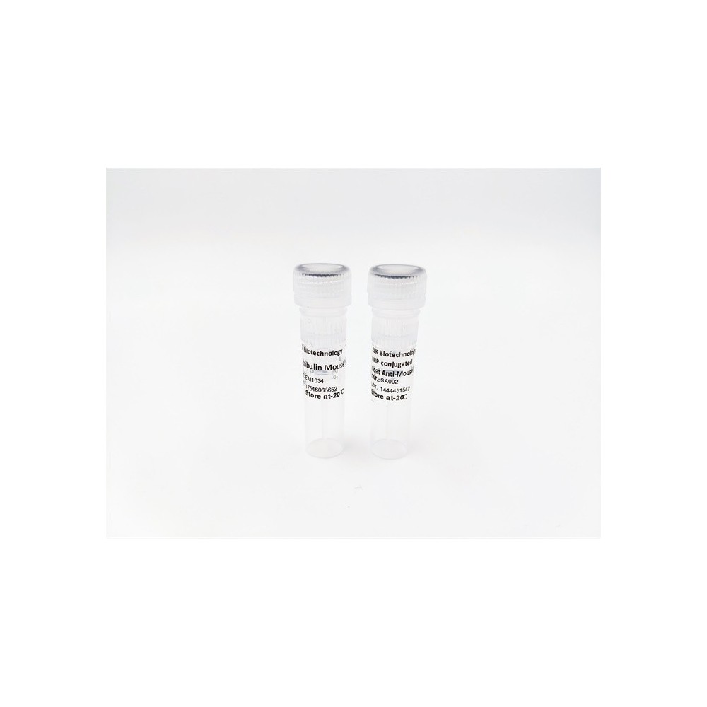

| ELK.No | ES6781 |
| Product name | PKC ε rabbit pAb |
| Reactivity | Human;Mouse;Rat |
| Applications | WB;ELISA;IHC |
| Other name | PRKCE; PKCE; Protein kinase C epsilon type; nPKC-epsilon |
| Size | 50μL |
| Unit price ($) | 148 |
| Human gene ID | 5581 |
| Human Swiss-Prot | Q02156 |
| Source | Rabbit |
| Isotype | IgG |
| Target | PKC ε |
| Fields | >>cGMP-PKG signaling pathway;>>Sphingolipid signaling pathway;>>Vascular smooth muscle contraction;>>Apelin signaling pathway;>>Tight junction;>>Fc gamma R-mediated phagocytosis;>>Inflammatory mediator regulation of TRP channels;>>Aldosterone synthesis and secretion;>>Type II diabetes mellitus;>>Insulin resistance;>>AGE-RAGE signaling pathway in diabetic complications;>>Shigellosis;>>MicroRNAs in cancer |
| Gene name | PRKCE |
| Protein name | Protein kinase C epsilon type |
| Human gene link | View Human Gene |
| Human Swiss link | View Human Swiss-Prot |
| Mouse gene ID | 18754 |
| Mouse gene link | View Mouse Gene |
| Mouse Swiss-Prot | P16054 |
| Mouse Swiss link | View Mouse Swiss-Prot |
| Rat gene ID | 29340 |
| Rat gene link | View Rat Gene |
| Rat Swiss-Prot | P09216 |
| Rat Swiss link | View Rat Swiss-Prot |
| Immunogen | The antiserum was produced against synthesized peptide derived from human PKC epsilon. AA range:688-737 |
| Specificity | PKC ε Polyclonal Antibody detects endogenous levels of PKC ε protein. |
| Formulation | Liquid in PBS containing 50% glycerol, 0.5% BSA and 0.02% sodium azide. |
| Clonality | Polyclonal |
| Dilution | WB 1:500-2000;IHC-p 1:50-300; ELISA 2000-20000 |
| Purification | The antibody was affinity-purified from rabbit antiserum by affinity-chromatography using epitope-specific immunogen. |
| Concentration | 1 mg/ml |
| Storage stability | -20°C/1 year |
| Molecular Weight (Da) | |
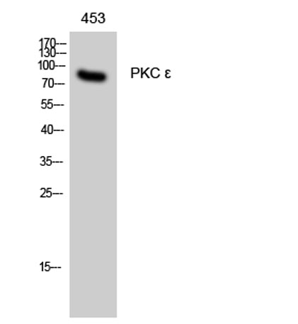
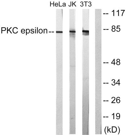
| Observed band (KD) | 83kD |
| Background | protein kinase C epsilon(PRKCE) Homo sapiens Protein kinase C (PKC) is a family of serine- and threonine-specific protein kinases that can be activated by calcium and the second messenger diacylglycerol. PKC family members phosphorylate a wide variety of protein targets and are known to be involved in diverse cellular signaling pathways. PKC family members also serve as major receptors for phorbol esters, a class of tumor promoters. Each member of the PKC family has a specific expression profile and is believed to play a distinct role in cells. The protein encoded by this gene is one of the PKC family members. This kinase has been shown to be involved in many different cellular functions, such as neuron channel activation, apoptosis, cardioprotection from ischemia, heat shock response, as well as insulin exocytosis. Knockout studies in mice suggest that this kinase is important for lipopolysaccharide (LPS)-mediated signaling in activated macro |
| Function | catalytic activity:ATP + a protein = ADP + a phosphoprotein.,domain:The C1 domain, containing the phorbol ester/DAG-type region 1 (C1A) and 2 (C1B), is the diacylglycerol sensor and the C2 domain is a non-calcium binding domain.,enzyme regulation:Three specific sites; Thr-566 (activation loop of the kinase domain), Thr-710 (turn motif) and Ser-729 (hydrophobic region), need to be phosphorylated for its full activation.,function:PKC is activated by diacylglycerol which in turn phosphorylates a range of cellular proteins. PKC also serves as the receptor for phorbol esters, a class of tumor promoters.,function:This is calcium-independent, phospholipid-dependent, serine- and threonine-specific enzyme.,PTM:Phosphorylation on Thr-566 by PDPK1 triggers autophosphorylation on Ser-729.,similarity:Belongs to the protein kinase superfamily. AGC Ser/Thr protein kinase family. PKC subfamily.,similari |
| Subcellular location | Cytoplasm . Cytoplasm, cytoskeleton . Cell membrane . Cytoplasm, perinuclear region . Nucleus . Translocated to plasma membrane in epithelial cells stimulated by HGF (PubMed:17603037). Associated with the Golgi at the perinuclear site in pre-passage fibroblasts (By similarity). In passaging cells, translocated to the cell periphery (By similarity). Translocated to the nucleus in PMA-treated cells (By similarity). . |
| Expression | Expressed in cumulus cells (at protein level). |

Use collapsible tabs for more detailed information that will help customers make a purchasing decision.
Ex: Shipping and return policies, size guides, and other common questions.