 Français
 English

| ELK.No | ES6910 |
| Product name | BAI-1 rabbit pAb |
| Reactivity | Human;Mouse |
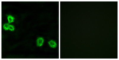
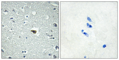
| Applications | WB;IHC;IF;ELISA |
| Other name | BAI1; Brain-specific angiogenesis inhibitor 1 |
| Size | 50μL |
| Unit price ($) | 148 |
| Human gene ID | 575 |
| Human Swiss-Prot | O14514 |
| Source | Rabbit |
| Isotype | IgG |
| Target | BAI-1 |
| Fields | >>p53 signaling pathway |
| Gene name | BAI1 |
| Protein name | Brain-specific angiogenesis inhibitor 1 |
| Human gene link | View Human Gene |
| Human Swiss link | View Human Swiss-Prot |
| Mouse gene ID | 107831 |
| Mouse gene link | View Mouse Gene |
| Mouse Swiss-Prot | Q3UHD1 |
| Mouse Swiss link | View Mouse Swiss-Prot |
| Rat gene ID | |
| Rat gene link | |
| Rat Swiss-Prot | |
| Rat Swiss link | |
| Immunogen | The antiserum was produced against synthesized peptide derived from human BAI1. AA range:691-740 |
| Specificity | BAI-1 Polyclonal Antibody detects endogenous levels of BAI-1 protein. |
| Formulation | Liquid in PBS containing 50% glycerol, 0.5% BSA and 0.02% sodium azide. |
| Clonality | Polyclonal |
| Dilution | Western Blot: 1/500 - 1/2000. Immunohistochemistry: 1/100 - 1/300. Immunofluorescence: 1/200 - 1/1000. ELISA: 1/10000. Not yet tested in other applications. |
| Purification | The antibody was affinity-purified from rabbit antiserum by affinity-chromatography using epitope-specific immunogen. |
| Concentration | 1 mg/ml |
| Storage stability | -20°C/1 year |
| Molecular Weight (Da) | |
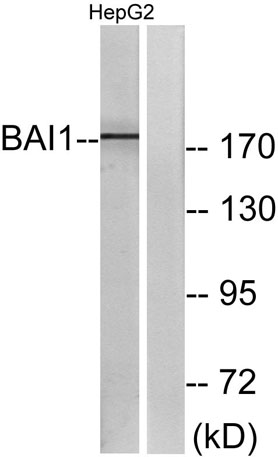
| Observed band (KD) | 174kD |
| Background | Angiogenesis is controlled by a local balance between stimulators and inhibitors of new vessel growth and is suppressed under normal physiologic conditions. Angiogenesis has been shown to be essential for growth and metastasis of solid tumors. In order to obtain blood supply for their growth, tumor cells are potently angiogenic and attract new vessels as results of increased secretion of inducers and decreased production of endogenous negative regulators. BAI1 contains at least one 'functional' p53-binding site within an intron, and its expression has been shown to be induced by wildtype p53. There are two other brain-specific angiogenesis inhibitor genes, designated BAI2 and BAI3 which along with BAI1 have similar tissue specificities and structures, however only BAI1 is transcriptionally regulated by p53. BAI1 is postulated to be a member of the secretin receptor family, |
| Function | domain:The TSP1 repeats inhibit in vivo angiogenesis in rat cornea induced by BFGF.,function:Phosphatidylserine receptor that enhances the engulfment of apoptotic cells. Likely to be a potent inhibitor of angiogenesis in brain and may play a significant role as a mediator of the p53 signal in suppression of glioblastoma. May function in cell adhesion and signal transduction in the brain.,induction:By p53.,similarity:Belongs to the G-protein coupled receptor 2 family. LN-TM7 subfamily.,similarity:Contains 1 GPS domain.,similarity:Contains 5 TSP type-1 domains.,subcellular location:Likely to be concentrated at cell-cell adhesion sites.,subunit:Interacts with ELMO1 and DOCK1. When bound to ELMO1 and DOCK1, it may act as a module to promote the engulfment (By similarity). Interacts with MAGI1, MAGI3, BAIAP2 and PHYHIP.,tissue specificity:Specifically expressed in brain. Reduced or no express |
| Subcellular location | Cell membrane ; Multi-pass membrane protein . Cell projection, phagocytic cup . Cell junction, focal adhesion . Cell projection, dendritic spine . Cell junction, synapse, postsynaptic density .; [Vasculostatin-120]: Secreted .; [Vasculostatin-40]: Secreted . |
| Expression | Expressed in brain (at protein level) (PubMed:12074842, PubMed:12507886). Expressed on mononuclear phagocytes and monocyte-derived macrophages in the gastric mucosa (at protein level) (PubMed:24509909). Expressed in normal pancreatic tissue but not in pancreatic tumor tissue (PubMed:11875720). Reduced or no expression is observed in some glioblastomas (PubMed:12507886). |

Use collapsible tabs for more detailed information that will help customers make a purchasing decision.
Ex: Shipping and return policies, size guides, and other common questions.