 Français
 English
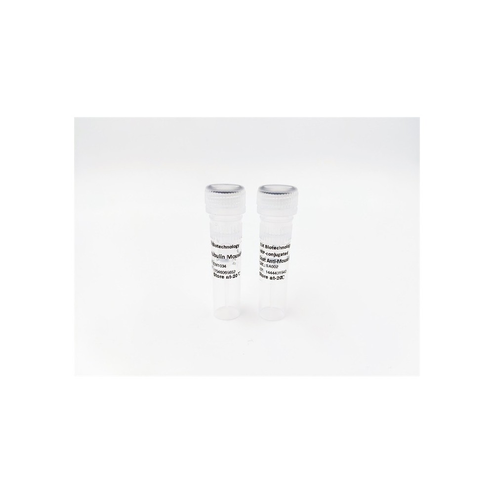

| ELK.No | ES7285 |
| Product name | SRPK1 rabbit pAb |
| Reactivity | Human;Mouse;Rat |
| Applications | WB;ELISA |
| Other name | SRPK1; SRSF protein kinase 1; SFRS protein kinase 1; Serine/arginine-rich protein-specific kinase 1; SR-protein-specific kinase 1 |
| Size | 50μL |
| Unit price ($) | 148 |
| Human gene ID | 6732 |
| Human Swiss-Prot | Q96SB4 |
| Source | Rabbit |
| Isotype | IgG |
| Target | SRPK1 |
| Fields | >>Herpes simplex virus 1 infection |
| Gene name | SRPK1 |
| Protein name | SRSF protein kinase 1 |
| Human gene link | View Human Gene |
| Human Swiss link | View Human Swiss-Prot |
| Mouse gene ID | 20815 |
| Mouse gene link | View Mouse Gene |
| Mouse Swiss-Prot | O70551 |
| Mouse Swiss link | View Mouse Swiss-Prot |
| Rat gene ID | |
| Rat gene link | |
| Rat Swiss-Prot | |
| Rat Swiss link | |
| Immunogen | The antiserum was produced against synthesized peptide derived from human SRPK1. AA range:521-570 |
| Specificity | SRPK1 Polyclonal Antibody detects endogenous levels of SRPK1 protein. |
| Formulation | Liquid in PBS containing 50% glycerol, 0.5% BSA and 0.02% sodium azide. |
| Clonality | Polyclonal |
| Dilution | Western Blot: 1/500 - 1/2000. ELISA: 1/40000. Not yet tested in other applications. |
| Purification | The antibody was affinity-purified from rabbit antiserum by affinity-chromatography using epitope-specific immunogen. |
| Concentration | 1 mg/ml |
| Storage stability | -20°C/1 year |
| Molecular Weight (Da) | |
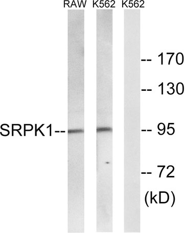
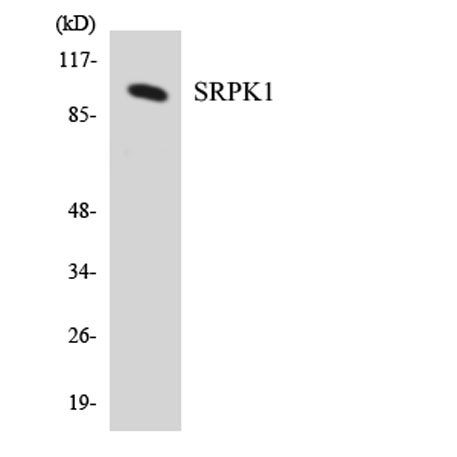
| Observed band (KD) | 95kD |
| Background | This gene encodes a serine/arginine protein kinase specific for the SR (serine/arginine-rich domain) family of splicing factors. The protein localizes to the nucleus and the cytoplasm. It is thought to play a role in regulation of both constitutive and alternative splicing by regulating intracellular localization of splicing factors. Alternative splicing of this gene results in multiple transcript variants. Additional alternatively spliced transcript variants have been described for this gene, but their full length nature have not been determined.[provided by RefSeq, Jul 2010], |
| Function | catalytic activity:ATP + a protein = ADP + a phosphoprotein.,cofactor:Magnesium.,enzyme regulation:Activated by phosphorylation on Ser-51 and Ser-555.,function:Plays a central role in the regulatory network for splicing, controlling the intranuclear distribution of splicing factors in interphase cells and the reorganization of nuclear speckles during mitosis. Hyperphosphorylates RS domain-containing proteins such as SFRS1 and SFRS2 on serine residues during metaphase but at lower levels during interphase. Locks onto SFRS1 to form a stable complex and processively phosphorylates the RS domain. Appears to mediate HBV core protein phosphorylation which is a prerequisite for pregenomic RNA encapsidation into viral capsids.,similarity:Belongs to the protein kinase superfamily. CMGC Ser/Thr protein kinase family.,similarity:Contains 1 protein kinase domain.,subunit:Present in a seven component |
| Subcellular location | [Isoform 2]: Cytoplasm. Nucleus. Nucleus matrix. Microsome. Shuttles between the nucleus and the cytoplasm. Inhibition of the Hsp90 ATPase activity, osmotic stress and interaction with HHV-1 ICP27 protein can induce its translocation to the nucleus. KAT5/TIP60 inhibits its nuclear translocation.; [Isoform 1]: Cytoplasm. Nucleus matrix. Microsome. Mainly localized in the microsomal fraction and the cytoplasm, and to a lesser extent in the nuclear matrix.; Cytoplasm . Nucleus, nucleoplasm . Nucleus speckle . Chromosome . Preferentially localizes to the promoter of gene coding regions. . |
| Expression | Isoform 2 is predominantly expressed in the testis but is also present at lower levels in heart, ovary, small intestine, liver, kidney, pancreas and skeletal muscle. Isoform 1 is only seen in the testis, at lower levels than isoform 2. Highly expressed in different erythroid and lymphoid cell lines, with isoform 2 being far more abundant than isoform 1. |

Use collapsible tabs for more detailed information that will help customers make a purchasing decision.
Ex: Shipping and return policies, size guides, and other common questions.