 Français
 English
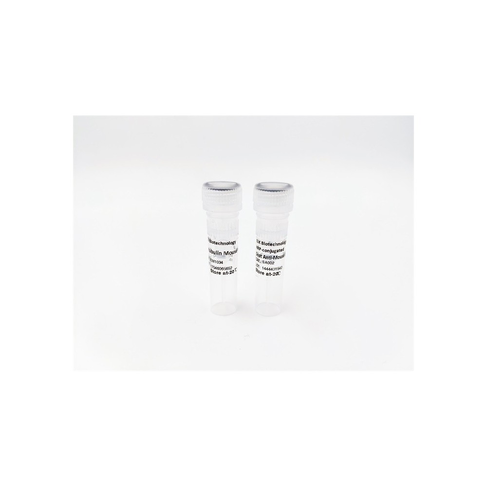
VCP (phospho Ser352) rabbit pAb
AO-06-ES7491-50

| ELK.No | ES7491 |
| Product name | VCP (phospho Ser352) rabbit pAb |
| Reactivity | Human;Mouse;Rat |
| Applications | WB;ELISA |
| Other name | VCP; Transitional endoplasmic reticulum ATPase; TER ATPase; 15S Mg(2+)-ATPase p97 subunit; Valosin-containing protein; VCP |
| Size | 50μL |
| Unit price ($) | 148 |
| Human gene ID | 7415 |
| Human Swiss-Prot | P55072 |
| Source | Rabbit |
| Isotype | IgG |
| Target | VCP |
| Fields | >>Protein processing in endoplasmic reticulum;>>Amyotrophic lateral sclerosis;>>Pathways of neurodegeneration - multiple diseases;>>Legionellosis |
| Gene name | VCP |
| Protein name | Transitional endoplasmic reticulum ATPase |
| Human gene link | View Human Gene |
| Human Swiss link | View Human Swiss-Prot |
| Mouse gene ID | 269523 |
| Mouse gene link | View Mouse Gene |
| Mouse Swiss-Prot | Q01853 |
| Mouse Swiss link | View Mouse Swiss-Prot |
| Rat gene ID | 116643 |
| Rat gene link | View Rat Gene |
| Rat Swiss-Prot | P46462 |
| Rat Swiss link | View Rat Swiss-Prot |
| Immunogen | The antiserum was produced against synthesized peptide derived from human VCP around the phosphorylation site of Ser352. AA range:318-367 |
| Specificity | Phospho-VCP (S352) Polyclonal Antibody detects endogenous levels of VCP protein only when phosphorylated at S352. |
| Formulation | Liquid in PBS containing 50% glycerol, 0.5% BSA and 0.02% sodium azide. |
| Clonality | Polyclonal |
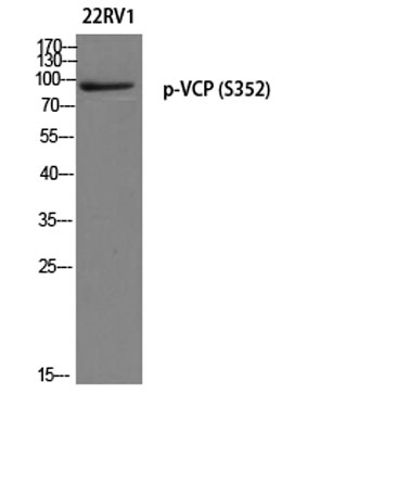
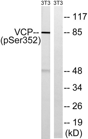
| Dilution | Western Blot: 1/500 - 1/2000. ELISA: 1/5000. Not yet tested in other applications. |
| Purification | The antibody was affinity-purified from rabbit antiserum by affinity-chromatography using epitope-specific immunogen. |
| Concentration | 1 mg/ml |
| Storage stability | -20°C/1 year |
| Molecular Weight (Da) | |
| Observed band (KD) | 85kD |
| Background | valosin containing protein(VCP) Homo sapiens The protein encoded by this gene is a member of a family that includes putative ATP-binding proteins involved in vesicle transport and fusion, 26S proteasome function, and assembly of peroxisomes. This protein, as a structural protein, is associated with clathrin, and heat-shock protein Hsc70, to form a complex. It has been implicated in a number of cellular events that are regulated during mitosis, including homotypic membrane fusion, spindle pole body function, and ubiquitin-dependent protein degradation. [provided by RefSeq, Jul 2008], |
| Function | disease:Defects in VCP are the cause of inclusion body myopathy with early-onset Paget disease and frontotemporal dementia (IBMPFD) [MIM:167320]; also known as muscular dystrophy, limb-girdle, with Paget disease of bone or pagetoid amyotrophic lateral sclerosis or pagetoid neuroskeletal syndrome or lower motor neuron degeneration with Paget-like bone disease. IBMPFD features adult-onset proximal and distal muscle weakness (clinically resembling limb girdle muscular dystrophy), early-onset Paget disease of bone in most cases and premature frontotemporal dementia.,function:Necessary for the fragmentation of Golgi stacks during mitosis and for their reassembly after mitosis. Involved in the formation of the transitional endoplasmic reticulum (tER). The transfer of membranes from the endoplasmic reticulum to the Golgi apparatus occurs via 50-70 nm transition vesicles which derive from part-r |
| Subcellular location | Cytoplasm, cytosol . Endoplasmic reticulum . Nucleus . Cytoplasm, Stress granule . Present in the neuronal hyaline inclusion bodies specifically found in motor neurons from amyotrophic lateral sclerosis patients (PubMed:15456787). Present in the Lewy bodies specifically found in neurons from Parkinson disease patients (PubMed:15456787). Recruited to the cytoplasmic surface of the endoplasmic reticulum via interaction with AMFR/gp78 (PubMed:16168377). Following DNA double-strand breaks, recruited to the sites of damage (PubMed:22120668). Recruited to stalled replication forks via interaction with SPRTN (PubMed:23042605). Recruited to damaged lysosomes decorated with K48-linked ubiquitin chains (PubMed:27753622). Colocalizes with TIA1, ZFAND1 and G3BP1 in cytoplasmic stress granules (SGs) in |
| Expression | Brain,Epithelium,Fetal brain cortex,Kidney,Lymph,PCR rescued clones,Pituitary,Plate |

Use collapsible tabs for more detailed information that will help customers make a purchasing decision.
Ex: Shipping and return policies, size guides, and other common questions.