 Français
 English

| ELK.No | ES7864 |
| Product name | Megsin rabbit pAb |
| Reactivity | Human;Rat;Mouse; |
| Applications | WB;ELISA |
| Other name | SERPINB7; Serpin B7; Megsin; TP55 |
| Size | 50μL |
| Unit price ($) | 148 |
| Human gene ID | 8710 |
| Human Swiss-Prot | O75635 |
| Source | Rabbit |
| Isotype | IgG |
| Target | Megsin |
| Fields | |
| Gene name | SERPINB7 |
| Protein name | Serpin B7 |
| Human gene link | View Human Gene |
| Human Swiss link | View Human Swiss-Prot |
| Mouse gene ID | |
| Mouse gene link | |
| Mouse Swiss-Prot | Q9D695 |
| Mouse Swiss link | View Mouse Swiss-Prot |
| Rat gene ID | |
| Rat gene link | |
| Rat Swiss-Prot | |
| Rat Swiss link | |
| Immunogen | The antiserum was produced against synthesized peptide derived from human SERPINB7. AA range:111-160 |
| Specificity | Megsin Polyclonal Antibody detects endogenous levels of Megsin protein. |
| Formulation | Liquid in PBS containing 50% glycerol, 0.5% BSA and 0.02% sodium azide. |
| Clonality | Polyclonal |
| Dilution | Western Blot: 1/500 - 1/2000. ELISA: 1/20000. Not yet tested in other applications. |
| Purification | The antibody was affinity-purified from rabbit antiserum by affinity-chromatography using epitope-specific immunogen. |
| Concentration | 1 mg/ml |
| Storage stability | -20°C/1 year |
| Molecular Weight (Da) | |
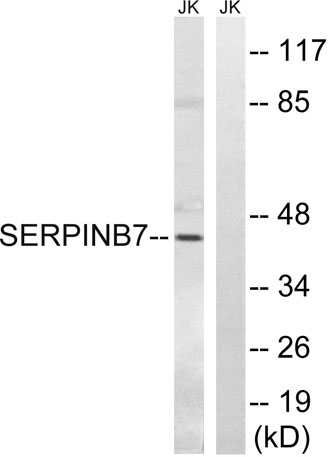
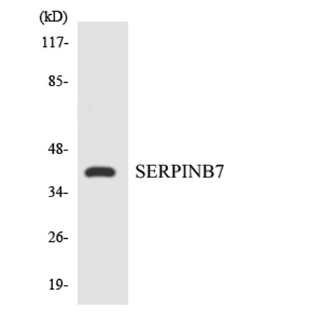
| Observed band (KD) | 43kD |
| Background | This gene encodes a member of a family of proteins which function as protease inhibitors. Expression of this gene is upregulated in IgA nephropathy and mutations have been found to cause palmoplantar keratoderma, Nagashima type. Alternative splicing results in multiple transcript variants. [provided by RefSeq, Jun 2014], |
| Function | function:Might function as an inhibitor of Lys-specific proteases. Might influence the maturation of megakaryocytes via its action as a serpin.,similarity:Belongs to the serpin family.,similarity:Belongs to the serpin family. Ov-serpin subfamily.,tissue specificity:Predominantly expressed in mesangial cells., |
| Subcellular location | Cytoplasm . |
| Expression | Predominantly expressed in mesangial cells. Expressed in the epidermis of the whole body. |

Use collapsible tabs for more detailed information that will help customers make a purchasing decision.
Ex: Shipping and return policies, size guides, and other common questions.