 Français
 English
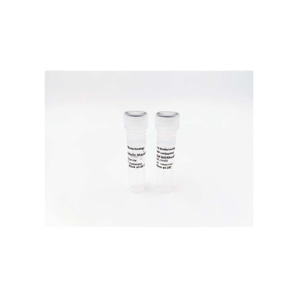

| ELK.No | ES8550 |
| Product name | COL17A1 rabbit pAb |
| Reactivity | Human;Rat;Mouse; |
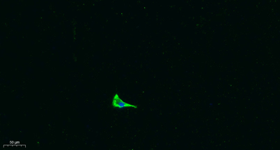
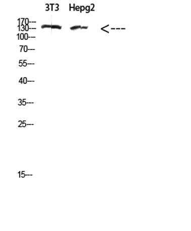
| Applications | WB IF;ELISA |
| Other name | COL17A1 BP180 BPAG2 |
| Size | 100μL |
| Unit price ($) | 248 |
| Human gene ID | 1308 |
| Human Swiss-Prot | Q9UMD9 |
| Source | Rabbit |
| Isotype | IgG |
| Target | COL17A1 |
| Fields | >>Protein digestion and absorption |
| Gene name | COL17A1 BP180 BPAG2 |
| Protein name | collagen, type XVII, alpha 1 |
| Human gene link | View Human Gene |
| Human Swiss link | View Human Swiss-Prot |
| Mouse gene ID | 12821 |
| Mouse gene link | |
| Mouse Swiss-Prot | Q07563 |
| Mouse Swiss link | View Mouse Swiss-Prot |
| Rat gene ID | |
| Rat gene link | |
| Rat Swiss-Prot | |
| Rat Swiss link | |
| Immunogen | The antiserum was produced against synthesized peptide derived from the Internal region of human COL17A1. AA range:481-530 |
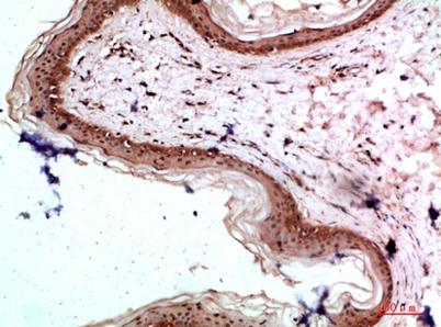
| Specificity | The antibody detects endogenous COL17A1 protein |
| Formulation | Liquid in PBS containing 50% glycerol, 0.5% BSA and 0.02% sodium azide. |
| Clonality | Polyclonal |
| Dilution | WB 1:500-2000 IF 1:100-300 ELISA 1:5000-20000 Not yet tested in other applications. |
| Purification | The antibody was affinity-purified from rabbit antiserum by affinity-chromatography using epitope-specific immunogen. |
| Concentration | 1 mg/ml |
| Storage stability | -20°C/1 year |
| Molecular Weight (Da) | |
| Observed band (KD) | 150kD |
| Background | This gene encodes the alpha chain of type XVII collagen. Unlike most collagens, collagen XVII is a transmembrane protein. Collagen XVII is a structural component of hemidesmosomes, multiprotein complexes at the dermal-epidermal basement membrane zone that mediate adhesion of keratinocytes to the underlying membrane. Mutations in this gene are associated with both generalized atrophic benign and junctional epidermolysis bullosa. Two homotrimeric forms of type XVII collagen exist. The full length form is the transmembrane protein. A soluble form, referred to as either ectodomain or LAD-1, is generated by proteolytic processing of the full length form. [provided by RefSeq, Jul 2008], |
| Function | disease:Defects in COL17A1 are a cause of generalized atrophic benign epidermolysis bullosa (GABEB) [MIM:226650]. GABEB is a non-lethal, adult form of junctional epidermolysis bullosa characterized by life-long blistering of the skin, associated with hair and tooth abnormalities.,function:May play a role in the integrity of hemidesmosome and the attachment of basal keratinocytes to the underlying basement membrane.,function:The 120 kDa linear IgA disease antigen is an anchoring filament component involved in dermal-epidermal cohesion. Is the target of linear IgA bullous dermatosis autoantibodies.,miscellaneous:Both the 120 kDa linear IgA disease antigen and the 97 kDa linear IgA disease antigen of COL17A1, represent major antigenic targets of autoantibodies in patients with linear IgA disease (LAD). LAD is a subepidermal blistering disorder characterized by tissue-bound and circulating I |
| Subcellular location | Cell junction, hemidesmosome. Membrane; Single-pass type II membrane protein. Localized along the plasma membrane of the hemidesmosome.; [120 kDa linear IgA disease antigen]: Secreted, extracellular space, extracellular matrix, basement membrane. Exclusively localized to anchoring filaments. Localized to the epidermal side of split skin.; [97 kDa linear IgA disease antigen]: Secreted, extracellular space, extracellular matrix, basement membrane. Localized in the lamina lucida beneath the hemidesmosomes. |
| Expression | Detected in skin (PubMed:8618013). In the cornea, it is detected in the epithelial basement membrane, the epithelial cells, and at a lower level in stromal cells (at protein level) (PubMed:25676728). Stratified squamous epithelia. Found in hemidesmosomes. Expressed in cornea, oral mucosa, esophagus, intestine, kidney collecting ducts, ureter, bladder, urethra and thymus but is absent in lung, blood vessels, skeletal muscle and nerves. |

Use collapsible tabs for more detailed information that will help customers make a purchasing decision.
Ex: Shipping and return policies, size guides, and other common questions.