 Français
 English

| ELK.No | ES8607 |
| Product name | GRP78 BiP rabbit pAb |
| Reactivity | Human;Mouse;Rat;Fish |
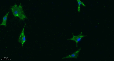
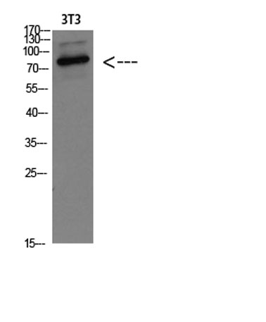
| Applications | WB;IHC;IF;ELISA |
| Other name | 78 kDa glucose-regulated protein (GRP-78) (Endoplasmic reticulum lumenal Ca(2+)-binding protein grp78) (Heat shock 70 kDa protein 5) (Immunoglobulin heavy chain-binding protein) (BiP) |
| Size | 100μL |
| Unit price ($) | 248 |
| Human gene ID | 3309 |
| Human Swiss-Prot | P11021 |
| Source | Rabbit |
| Isotype | IgG |
| Target | HSP A5/GRP78 |
| Fields | >>Protein export;>>Protein processing in endoplasmic reticulum;>>Antigen processing and presentation;>>Thyroid hormone synthesis;>>Parkinson disease;>>Amyotrophic lateral sclerosis;>>Prion disease;>>Pathways of neurodegeneration - multiple diseases;>>Lipid and atherosclerosis |
| Gene name | HSPA5 GRP78 |
| Protein name | GRP78 BiP |
| Human gene link | View Human Gene |
| Human Swiss link | View Human Swiss-Prot |
| Mouse gene ID | 14828 |
| Mouse gene link | |
| Mouse Swiss-Prot | P20029 |
| Mouse Swiss link | View Mouse Swiss-Prot |
| Rat gene ID | |
| Rat gene link | |
| Rat Swiss-Prot | P06761 |
| Rat Swiss link | View Rat Swiss-Prot |
| Immunogen | Synthetic peptide from human protein at AA range: 505-570 |
| Specificity | The antibody detects endogenous GRP78 BiP |
| Formulation | Liquid in PBS containing 50% glycerol, 0.5% BSA and 0.02% sodium azide. |
| Clonality | Polyclonal |
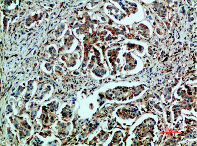
| Dilution | Western Blot: 1/500 - 1/2000.IHC-p/f:1:50-300. ELISA 1:10000-20000 |
| Purification | The antibody was affinity-purified from rabbit antiserum by affinity-chromatography using epitope-specific immunogen. |
| Concentration | 1 mg/ml |
| Storage stability | -20°C/1 year |
| Molecular Weight (Da) | |
| Observed band (KD) | 78kD |
| Background | The protein encoded by this gene is a member of the heat shock protein 70 (HSP70) family. It is localized in the lumen of the endoplasmic reticulum (ER), and is involved in the folding and assembly of proteins in the ER. As this protein interacts with many ER proteins, it may play a key role in monitoring protein transport through the cell.[provided by RefSeq, Sep 2010], |
| Function | disease:Autoantigen in rheumatoid arthritis [MIM:180300].,function:Probably plays a role in facilitating the assembly of multimeric protein complexes inside the ER.,similarity:Belongs to the heat shock protein 70 family.,subcellular location:Identified by mass spectrometry in melanosome fractions from stage I to stage IV.,subunit:Interacts with DNAJC1 (via J domain) (By similarity). Component of an EIF2 complex at least composed of CUGBP1, CALR, CALR3, EIF2S1, EIF2S2, HSP90B1 and HSPA5. Part a large chaperone multiprotein complex comprising CABP1, DNAJB11, HSP90B1, HSPA5, HYOU, PDIA2, PDIA4, PPIB, SDF2L1, UGT1A1 and very small amounts of ERP29, but not, or at very low levels, CALR nor CANX. Interacts with TMEM132A., |
| Subcellular location | Endoplasmic reticulum lumen . Melanosome . Cytoplasm . Cell surface . Identified by mass spectrometry in melanosome fractions from stage I to stage IV (PubMed:12643545). Localizes to the cell surface of epithelial cells in response to high levels of free iron (PubMed:20484814, PubMed:24355926, PubMed:27159390). . |
| Expression | Articular cartilage,Brain,Cajal-Retzius cell,Cervix carcino |

Use collapsible tabs for more detailed information that will help customers make a purchasing decision.
Ex: Shipping and return policies, size guides, and other common questions.