 Français
 English
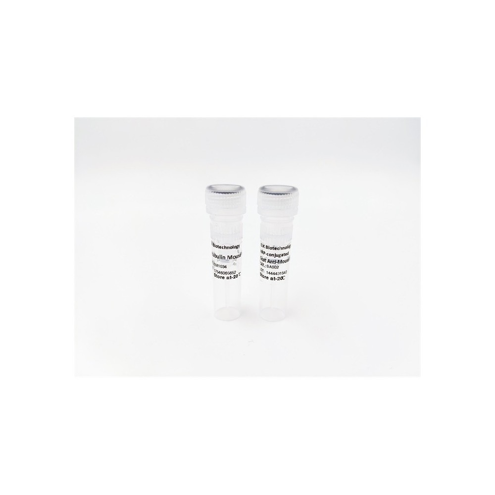

| ELK.No | ES8892 |
| Product name | TIE2 rabbit pAb |
| Reactivity | Human;Mouse;Rat |
| Applications | WB;IHC;IF;ELISA |
| Other name | Angiopoietin-1 receptor (EC 2.7.10.1;Endothelial tyrosine kinase;Tunica interna endothelial cell kinase;Tyrosine kinase with Ig and EGF homology domains-2;Tyrosine-protein kinase receptor TEK;Tyrosine-protein kinase receptor TIE-2;hTIE2;p140 TEK;CD antigen CD202b) |
| Size | 50μL |
| Unit price ($) | 148 |
| Human gene ID | 7010 |
| Human Swiss-Prot | Q02763 |
| Source | Rabbit |
| Isotype | IgG |
| Target | Tie-2 |
| Fields | >>MAPK signaling pathway;>>Ras signaling pathway;>>Rap1 signaling pathway;>>HIF-1 signaling pathway;>>PI3K-Akt signaling pathway;>>Rheumatoid arthritis |
| Gene name | TEK TIE2 VMCM VMCM1 |
| Protein name | Angiopoietin-1 receptor (EC 2.7.10.1) (Endothelial tyrosine kinase) (Tunica interna endothelial cell kinase) (Tyrosine kinase with Ig and EGF homology domains-2) (Tyrosine-protein kinase receptor TEK) |
| Human gene link | View Human Gene |
| Human Swiss link | View Human Swiss-Prot |
| Mouse gene ID | |
| Mouse gene link | |
| Mouse Swiss-Prot | Q02858 |
| Mouse Swiss link | View Mouse Swiss-Prot |
| Rat gene ID | |
| Rat gene link | |
| Rat Swiss-Prot | |
| Rat Swiss link | |
| Immunogen | Synthesized peptide derived from human TIE2 Polyclonal |
| Specificity | This antibody detects endogenous levels of TIE2. |
| Formulation | Liquid in PBS containing 50% glycerol, 0.5% BSA and 0.02% sodium azide. |
| Clonality | Polyclonal |
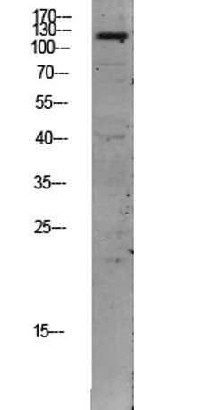
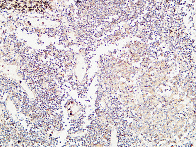
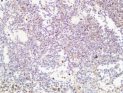
| Dilution | IHC-p: 100-300.WB 1:500-2000, ELISA 1:10000-20000 |
| Purification | The antibody was affinity-purified from rabbit antiserum by affinity-chromatography using epitope-specific immunogen. |
| Concentration | 1 mg/ml |
| Storage stability | -20°C/1 year |
| Molecular Weight (Da) | |
| Observed band (KD) | 120kD |
| Background | This gene encodes a receptor that belongs to the protein tyrosine kinase Tie2 family. The encoded protein possesses a unique extracellular region that contains two immunoglobulin-like domains, three epidermal growth factor (EGF)-like domains and three fibronectin type III repeats. The ligand angiopoietin-1 binds to this receptor and mediates a signaling pathway that functions in embryonic vascular development. Mutations in this gene are associated with inherited venous malformations of the skin and mucous membranes. Alternative splicing results in multiple transcript variants. Additional alternatively spliced transcript variants of this gene have been described, but their full-length nature is not known. [provided by RefSeq, Feb 2014], |
| Function | catalytic activity:ATP + a [protein]-L-tyrosine = ADP + a [protein]-L-tyrosine phosphate.,disease:Defects in TEK are a cause of dominantly inherited venous malformations (VMCM) [MIM:600195]; an error of vascular morphogenesis characterized by dilated, serpiginous channels.,function:This protein is a protein tyrosine-kinase transmembrane receptor for angiopoietin 1. It may constitute the earliest mammalian endothelial cell lineage marker. Probably regulates endothelial cell proliferation, differentiation and guides the proper patterning of endothelial cells during blood vessel formation.,similarity:Belongs to the protein kinase superfamily. Tyr protein kinase family.,similarity:Belongs to the protein kinase superfamily. Tyr protein kinase family. Tie subfamily.,similarity:Contains 1 protein kinase domain.,similarity:Contains 2 Ig-like C2-type (immunoglobulin-like) domains.,similarity:Cont |
| Subcellular location | Cell membrane ; Single-pass type I membrane protein. Cell junction . Cell junction, focal adhesion . Cytoplasm, cytoskeleton. Secreted . Recruited to cell-cell contacts in quiescent endothelial cells (PubMed:18425120, PubMed:18425119). Colocalizes with the actin cytoskeleton and at actin stress fibers during cell spreading. Recruited to the lower surface of migrating cells, especially the rear end of the cell. Proteolytic processing gives rise to a soluble extracellular domain that is secreted (PubMed:11806244). . |
| Expression | Detected in umbilical vein endothelial cells. Proteolytic processing gives rise to a soluble extracellular domain that is detected in blood plasma (at protein level). Predominantly expressed in endothelial cells and their progenitors, the angioblasts. Has been directly found in placenta and lung, with a lower level in umbilical vein endothelial cells, brain and kidney. |

Use collapsible tabs for more detailed information that will help customers make a purchasing decision.
Ex: Shipping and return policies, size guides, and other common questions.