 Français
 English
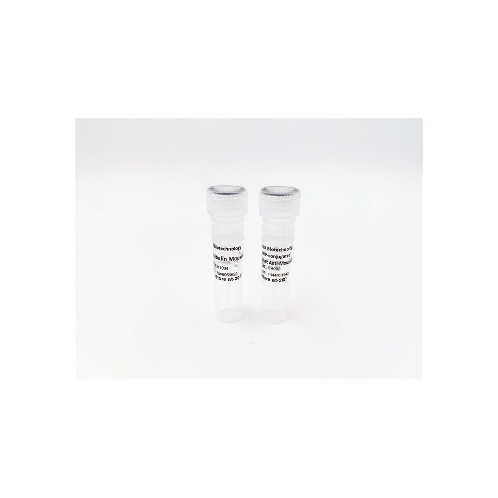

| ELK.No | ES8948 |
| Product name | PLSL rabbit pAb |
| Reactivity | Human;Mouse |
| Applications | WB;ELISA |
| Other name | |
| Size | 50μL |
| Unit price ($) | 148 |
| Human gene ID | 3936 |
| Human Swiss-Prot | P13796 |
| Source | Rabbit |
| Isotype | IgG |
| Target | PLSL |
| Fields | |
| Gene name | LCP1 PLS2 |
| Protein name | Plastin-2 (L-plastin) (LC64P) (Lymphocyte cytosolic protein 1) (LCP-1) |
| Human gene link | |
| Human Swiss link | View Human Swiss-Prot |
| Mouse gene ID | |
| Mouse gene link | |
| Mouse Swiss-Prot | Q61233 |
| Mouse Swiss link | View Mouse Swiss-Prot |
| Rat gene ID | |
| Rat gene link | |
| Rat Swiss-Prot | |
| Rat Swiss link | |
| Immunogen | Synthesized peptide derived from human protein . at AA range: 1-80 |
| Specificity | PLSL Polyclonal Antibody detects endogenous levels of protein. |
| Formulation | Liquid in PBS containing 50% glycerol, and 0.02% sodium azide. |
| Clonality | Polyclonal |
| Dilution | WB 1:500-2000 ELISA 1:5000-20000 |
| Purification | The antibody was affinity-purified from rabbit antiserum by affinity-chromatography using epitope-specific immunogen. |
| Concentration | 1 mg/ml |
| Storage stability | -20°C/1 year |
| Molecular Weight (Da) | |
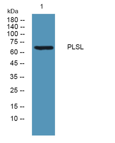
| Observed band (KD) | 68kD |
| Background | Plastins are a family of actin-binding proteins that are conserved throughout eukaryote evolution and expressed in most tissues of higher eukaryotes. In humans, two ubiquitous plastin isoforms (L and T) have been identified. Plastin 1 (otherwise known as Fimbrin) is a third distinct plastin isoform which is specifically expressed at high levels in the small intestine. The L isoform is expressed only in hemopoietic cell lineages, while the T isoform has been found in all other normal cells of solid tissues that have replicative potential (fibroblasts, endothelial cells, epithelial cells, melanocytes, etc.). However, L-plastin has been found in many types of malignant human cells of non-hemopoietic origin suggesting that its expression is induced accompanying tumorigenesis in solid tissues. [provided by RefSeq, Jul 2008], |
| Function | function:Actin-binding protein found in intestinal microvilli, hair cell stereocilia, and fibroblast filopodia.,PTM:Phosphorylated.,PTM:The N-terminus is blocked.,similarity:Contains 2 actin-binding domains.,similarity:Contains 2 EF-hand domains.,similarity:Contains 4 CH (calponin-homology) domains.,subunit:Monomer.,tissue specificity:Restricted to the spleen and other lymph node-containing organs. Expressed in neutrophils, monocytes, B lymphocytes, and myeloid cells., |
| Subcellular location | Cytoplasm, cytoskeleton . Cell junction . Cell projection . Cell projection, ruffle membrane ; Peripheral membrane protein ; Cytoplasmic side . Relocalizes to the immunological synapse between peripheral blood T-lymphocytes and antibody-presenting cells in response to costimulation through TCR/CD3 and CD2 or CD28 (PubMed:17294403). Associated with the actin cytoskeleton at membrane ruffles. Relocalizes to actin-rich cell projections upon serine phosphorylation (PubMed:16636079). . |
| Expression | Detected in intestinal microvilli, hair cell stereocilia, and fibroblast filopodia, in spleen and other lymph node-containing organs. Expressed in peripheral blood T-lymphocytes, neutrophils, monocytes, B-lymphocytes, and myeloid cells. |

Use collapsible tabs for more detailed information that will help customers make a purchasing decision.
Ex: Shipping and return policies, size guides, and other common questions.