 Français
 English

| ELK.No | ES9005 |
| Product name | NDRG1 rabbit pAb |
| Reactivity | Human;Mouse;Rat |
| Applications | WB;ELISA |
| Other name | |
| Size | 50μL |
| Unit price ($) | 148 |
| Human gene ID | 10397 |
| Human Swiss-Prot | Q92597 |
| Source | Rabbit |
| Isotype | IgG |
| Target | NDRG1 |
| Fields | |
| Gene name | NDRG1 CAP43 DRG1 RTP |
| Protein name | Protein NDRG1 (Differentiation-related gene 1 protein) (DRG-1) (N-myc downstream-regulated gene 1 protein) (Nickel-specific induction protein Cap43) (Reducing agents and tunicamycin-responsive protein |
| Human gene link | |
| Human Swiss link | View Human Swiss-Prot |
| Mouse gene ID | |
| Mouse gene link | |
| Mouse Swiss-Prot | Q62433 |
| Mouse Swiss link | View Mouse Swiss-Prot |
| Rat gene ID | |
| Rat gene link | |
| Rat Swiss-Prot | Q6JE36 |
| Rat Swiss link | View Rat Swiss-Prot |
| Immunogen | Synthesized peptide derived from human protein . at AA range: 270-350 |
| Specificity | NDRG1 Polyclonal Antibody detects endogenous levels of protein. |
| Formulation | Liquid in PBS containing 50% glycerol, and 0.02% sodium azide. |
| Clonality | Polyclonal |
| Dilution | WB 1:500-2000 ELISA 1:5000-20000 |
| Purification | The antibody was affinity-purified from rabbit antiserum by affinity-chromatography using epitope-specific immunogen. |
| Concentration | 1 mg/ml |
| Storage stability | -20°C/1 year |
| Molecular Weight (Da) | |
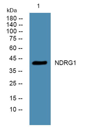
| Observed band (KD) | 43kD |
| Background | This gene is a member of the N-myc downregulated gene family which belongs to the alpha/beta hydrolase superfamily. The protein encoded by this gene is a cytoplasmic protein involved in stress responses, hormone responses, cell growth, and differentiation. The encoded protein is necessary for p53-mediated caspase activation and apoptosis. Mutations in this gene are a cause of Charcot-Marie-Tooth disease type 4D, and expression of this gene may be a prognostic indicator for several types of cancer. Alternatively spliced transcript variants encoding multiple isoforms have been observed for this gene. [provided by RefSeq, May 2012], |
| Function | disease:Defects in NDRG1 are the cause of Charcot-Marie-Tooth disease type 4D (CMT4D) [MIM:601455]; also known as hereditary motor and sensory neuropathy Lom type (HMSNL). CMT4D is a recessive form of Charcot-Marie-Tooth disease, the most common inherited disorder of the peripheral nervous system. Charcot-Marie-Tooth disease is classified in two main groups on the basis of electrophysiologic properties and histopathology: primary peripheral demyelinating neuropathy and primary peripheral axonal neuropathy. Demyelinating CMT neuropathies are characterized by severely reduced nerve conduction velocities (less than 38 m/sec), segmental demyelination and remyelination with onion bulb formations on nerve biopsy, slowly progressive distal muscle atrophy and weakness, absent deep tendon reflexes, and hollow feet. By convention, autosomal recessive forms of demyelinating Charcot-Marie-Tooth dise |
| Subcellular location | Cytoplasm, cytosol. Cytoplasm, cytoskeleton, microtubule organizing center, centrosome. Nucleus. Cell membrane. Mainly cytoplasmic but differentially localized to other regions. Associates with the plasma membrane in intestinal epithelia and lactating mammary gland. Translocated to the nucleus in a p53/TP53-dependent manner. In prostate epithelium and placental chorion, located in both the cytoplasm and in the nucleus. No nuclear localization in colon epithelium cells. In intestinal mucosa, prostate and renal cortex, located predominantly adjacent to adherens junctions. Cytoplasmic with granular staining in proximal tubular cells of the kidney and salivary gland ducts. Recruits to the membrane of recycling/sorting and late endosomes via binding to phosphatidylinositol 4-phosphate. Associat |
| Expression | Ubiquitous; expressed most prominently in placental membranes and prostate, kidney, small intestine, and ovary tissues. Also expressed in heart, brain, skeletal muscle, lung, liver and pancreas. Low levels in peripheral blood leukocytes and in tissues of the immune system. Expressed mainly in epithelial cells. Also found in Schwann cells of peripheral neurons. Reduced expression in adenocarcinomas compared to normal tissues. In colon, prostate and placental membranes, the cells that border the lumen show the highest expression. |

Use collapsible tabs for more detailed information that will help customers make a purchasing decision.
Ex: Shipping and return policies, size guides, and other common questions.