|

|
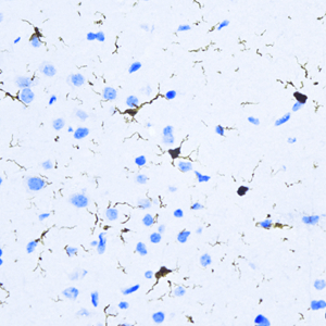
IHC analysis of IBA-1 (GB12105).
Sample: Mouse brain (Paraffin), 4% PFA (G1101) 12-24h.
Antigen retrieval: TE buffer (pH 9.0) (G1203),98℃,20min.
Blocking buffer: 3% BSA in PBS (GC305010), RT, 30min.
Primary antibody: 1: 2000, 4℃ overnight.
Secondary antibody: HRP Goat Anti-Mouse lgG (GB23301), 1: 200 RT 1h.
|
|

|
IHC analysis of IBA-1 (GB12105).
Sample: Rat brain (Paraffin), 4% PFA (G1101) 12-24h.
Antigen retrieval: TE buffer (pH 9.0) (G1203),98℃,20min.
Blocking buffer: 3% BSA in PBS (GC305010), RT, 30min.
Primary antibody: 1: 2000, 4℃ overnight.
Secondary antibody: HRP Goat Anti-Mouse lgG (GB23301), 1: 200 RT 1h.
|
 |
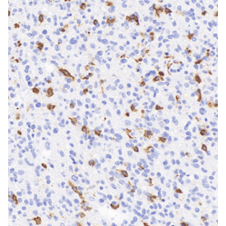
Immunohistochemistry of paraffin embedded rat brain using IBA-1 (GB12105) at dilution of 1: 2000 (300x lens)
|
|
|
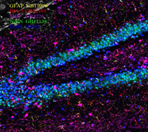
Immunofluorescent analysis of paraffin embedded rat hippocampus using Neun (GB11138) (green) + Iba1 (GB12105) (red) + MBP (GB12226) (spred) + GFAP (GB11096) (yellow) and Dapi in blue at dilution of 1: 1000
|
|

|
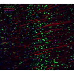
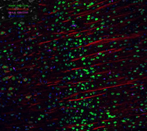
Immunofluorescence of paraffin embedded mouse brain using IBA-1 (GB12105) at dilution of 1: 1000 (200x lens)
|
|
|
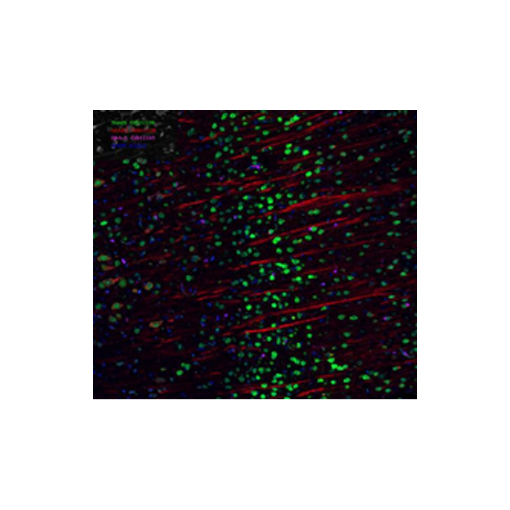
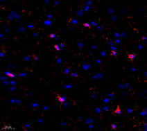
Immunofluorescent analysis of paraffin embedded rat brain using Neun (GB11138) (green)+ Iba1 (GB12105) (spred) + MAP2 (GB11128-2) (red) and Dapi in blue at dilution of 1: 1000
|
|
|
IF analysis of IBA-1 (GB12105).
Sample: Rat brain (Paraffin), 4% PFA (G1101) 12-24h.
Antigen retrieval: TE buffer (pH 9.0) (G1203),98℃,20min.
Blocking buffer: 3% BSA in PBS (GC305010), RT, 30min.
Primary antibody: 1: 1000, 4℃ overnight.
Secondary antibody: Cy3 conjugated Goat Anti-mouse IgG (H+L)(GB21301),1: 200 RT 1h.
|
|
|
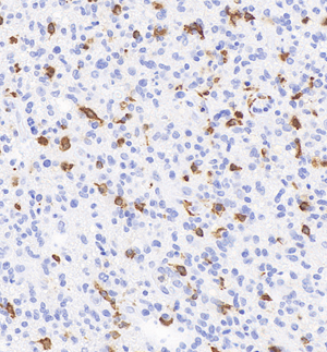
IHC analysis of IBA-1 (GB12105).
Sample: Human glioblastoma multiforme, 4% PFA (G1101) 12-24h.
Antigen retrieval: TE buffer (pH 9.0) (G1203),98℃,20min.
Blocking buffer: 3% BSA in PBS (GC305010), RT, 30min.
Primary antibody: 1: 500, 4℃ overnight.
Secondary antibody: S-vision poly-HRP conjugated Goat Anti-Rabbit lgG(H+L), Ready to use(G1302), RT, 20min.
|
|
|
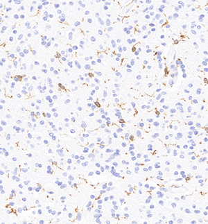
IHC analysis of IBA-1 (GB12105).
Sample: Human oligodendrocytoma , 4% PFA (G1101) 12-24h.
Antigen retrieval: TE buffer (pH 9.0) (G1203),98℃,20min.
Blocking buffer: 3% BSA in PBS (GC305010), RT, 30min.
Primary antibody: 1: 500, 4℃ overnight.
Secondary antibody: S-vision poly-HRP conjugated Goat Anti-Rabbit lgG(H+L), Ready to use(G1302), RT, 20min.
|
|
|
IHC analysis of IBA-1 (GB12105).
Sample: Monkey brain, 4% PFA (G1101) 12-24h.
Antigen retrieval: TE buffer (pH 9.0) (G1203),98℃,20min.
Blocking buffer: 3% BSA in PBS (GC305010), RT, 30min.
Primary antibody: 1: 500, 4℃ overnight.
Secondary antibody: S-vision poly-HRP conjugated Goat Anti-Rabbit lgG(H+L), Ready to use(G1302), RT, 20min.
|