
Annexin V-EGFP/PI Cell Apoptosis Detection Kit (Green EGFP + Red PI) 50T
AO-03-G1510-50T

Product Information
|
Product Name |
Cat. No |
Spec. |
|
Annexin V-EGFP/PI Cell Apoptosis Detection Kit (Green EGFP + Red PI) |
AO-03-G1510-50T |
50 T |
|
AO-03-G1510-100T |
100 T |
Product Description/Introduction
Apoptosis is a normal physiological process that occurs during embryonic development and maintenance of tissue homeostasis and is accompanied by many morphological changes, among which the loss of cell membrane is one of the early characteristics of apoptosis. In normal cells, phosphatidylserine (PS) is only distributed on the inner side of the phospholipid bilayer of the cell membrane. However, in the early stage of apoptosis, PS flips from the inner side of the lipid membrane to the outer side, exposing it to the outside of the cell. Annexin V is a Ca2+-dependent phospholipid-binding protein with high affinity for PS and it can specifically bind to cells exposed to PS. Therefore, Annexin V is used as one of the indicators to detect early apoptosis of cells. Propidium iodide (PI) is a nucleic acid dye that cannot penetrate normal cells with intact cell membranes and early apoptotic cells, but it can penetrate the cell membranes of late apoptotic and necrotic cells and stain cell nuclei.
This product uses a fusion protein composed of EGFP (enhanced Green Fluorescent Protein) and Annexin V as a detection probe to detect early apoptosis of cells. The PI is also used to distinguish live cells from necrotic and late apoptotic cells. In combination with Annexin V-EGFP and PI, live cells show negative staining (Annexin V-/PI-), early apoptotic cells show single fluorescence positive (Annexin V+/PI-), while late apoptotic and necrotic cells show double fluorescence positive (Annexin V+/PI+). This kit is suitable for flow cytometry or fluorescence microscopy detection. It is also suitable for quantitative detection of apoptotic cells as EGFP is fused 1:1 to Annexin V.
Storage and Shipping Conditions
Ship with wet ice; Store at 2-8℃ away from light, valid for 12 months.
Product Components
|
Component Number |
Component |
G1510-50T |
G1510-100T |
|
AO-03-G1510-1 |
Annexin V-EGFP |
250 μL |
2×250 μL |
|
AO-03-G1510-2 |
Propidium Iodide(PI) |
250 μL |
2×250 μL |
|
AO-03G1510-3 |
1×Binding Buffer |
25 mL |
2×25 mL |
|
Manual |
1 pc |
||
Product Protocol/Procedures
1. Suspension of cells: Take cell suspension and collect the cells by centrifugation at 500 x g for 5 min at 4℃.
Adherent cells: Collect cells culture supernatant first. then digest cells with trypsin without EDTA (recommended G4002 or G4011), combine with the cell culture supernatant and collect the cells by centrifugation at 500 x g for 5 min at 4℃. Trypsin digestion should not be too long to avoid false positives caused by excessive digestion.
2. Wash the cells twice with pre-cooled PBS (recommended G4202), and collect the cells by centrifugation at 500 x g for 5 min at 4℃ each time.
3. Gently resuspend the cells with pre-cooled 1×Binding Buffer, and adjust the cell concentration to 1~5×10⁶/mL.
4. Add 5 μL of Annexin V-EGFP and 5 μL of PI to 100 μL of cell suspension, mix gently and protect from light at room temperature for 8-10 min.
5. Add 400 µL of pre-cooled 1×Binding Buffer, shake gently, and use flow cytometry or fluorescence microscope for detection within 1 hour.
Result Analysis
1. Flow Cytometry Detection
a) Select the appropriate voltage and adjust the light compensation for the flow cytometer analysis. it is recommended to set a negative control (without Annexin V-EGFP and PI labeling) to adjust the voltage except for the experimental group, and the single standard control (with Annexin V-EGFP only, and cells with PI only) for compensation adjustment.
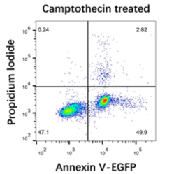
b) Reference example of flow cytometry detection and analysis: Induce Jurkat T lymphoma cells with 5 µM Camptothecin for 6 h. Referring to the above experimental steps, use flow cytometry to detect. The results are shown in the following figure.
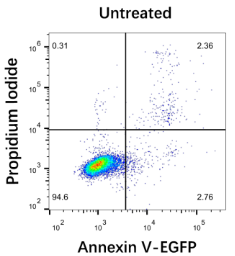
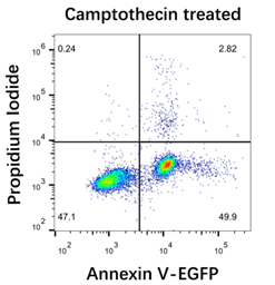
The maximum excitation wavelength of EGFP is 488 nm and the maximum emission wavelength is 507 nm; the maximum excitation wavelength of PI-DNA complex is 535 nm and the maximum emission wavelength is 615 nm. A two-colour scatter plot is plotted by flow cytometry correlation analysis software, with EGFP in the horizontal coordinate and PI in the vertical coordinate. In a typical experiment, live cells are non-fluorescent and the scatter point is located in the lower left first quadrant. Cells in early apoptosis have a strong green fluorescence and the scatter is in the second lower right quadrant. Late stage apoptotic and necrotic cells show dual red and green fluorescence, with the scatter point located in the upper right third quadrant.
2. Fluorescence Microscopy Detection
a) Add 5-10 µL of Annexin V-EGFP and PI double-stained cell suspension to the slide.
b) Cover with a coverslip.
c) Observe with a two-color filter under a fluorescence microscope. Annexin V-EGFP has a green fluorescence signal, and PI has a red fluorescence signal (When taking pictures with a fluorescence microscope, it is recommended to add an appropriate amount of anti-fluorescence quenching sealer (G1401) to prevent fluorescence quenching problems).
Note
1. The entire experimental process should be handled as gently as possible to avoid cell fragmentation, which may affect the experimental results.
2. Washing the cells with PBS cannot be omitted, and the residual PBS should also be removed as much as possible.
3. When using trypsin to digest cells, the experiment should be handled carefully and the digestion time should be controlled to avoid artificial damage to the cells. If the digestion time is too short, the cells need to be vigorously beaten to fall off, which may easily cause mechanical damage to the cell membrane; if the digestion time is too long, the cell membrane is also easily damaged and the results will be affected. In addition, trypsin containing EDTA cannot be used. EDTA will affect the binding of Annexin V to PS.
4. If some cells are floating after apoptosis stimulation, collect both the cell culture supernatant and the adherent cells to stain for a more accurate result.
5. Annexin V-EGFP and PI are sensitive to light, please avoid light during operation. Testing should be performed as soon as possible after the reaction is complete.
6. For your safety and health, please wear safety glasses, gloves, or protective clothing.
For Research Use Only!
Use collapsible tabs for more detailed information that will help customers make a purchasing decision.
Ex: Shipping and return policies, size guides, and other common questions.
