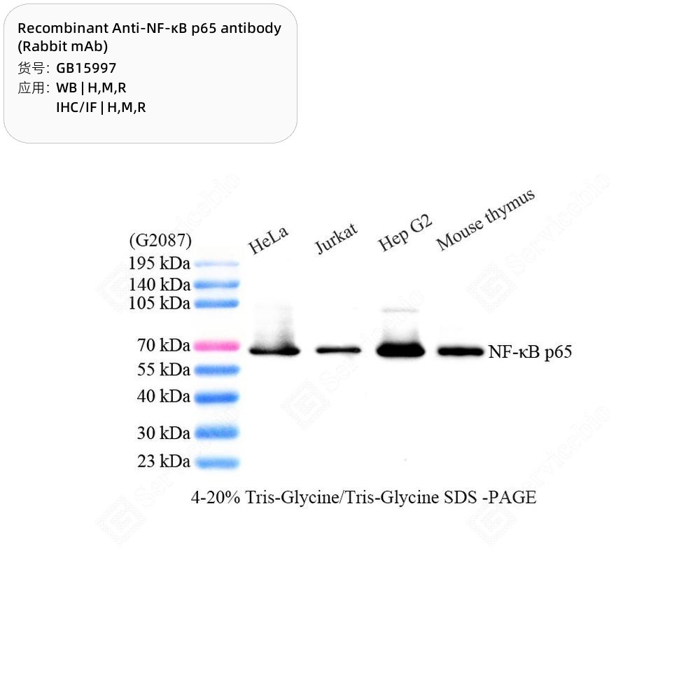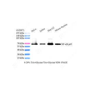
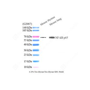
| WB analysis of NF-κB p65(GB15997).
Sample: Protein treated by RIPA Lysis Buffer (G2002).
Blocking buffer: 3% Nonfat dry milk (GC310001) in TBST, RT, 1h.
Primary antibody: 1: 1000, 4°C overnight.
Secondary antibody: HRP conjugated Goat Anti-Rabbit lgG (H+L) (GB23303), 1: 5000, RT, 1h. |
| WB analysis of NF-κB p65(GB15997).
Sample: Protein treated by RIPA Lysis Buffer (G2002).
Blocking buffer: 3% Nonfat dry milk (GC310001) in TBST, RT, 1h.
Primary antibody: 1: 1000, 4°C overnight.
Secondary antibody: HRP conjugated Goat Anti-Rabbit lgG (H+L) (GB23303), 1: 5000, RT, 1h. |
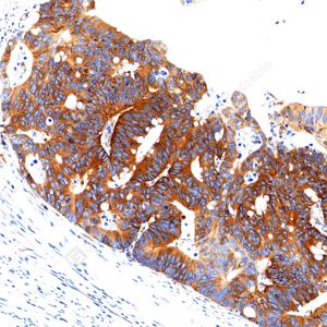
| IHC analysis of NF-κB p65(GB15997).
Sample: Human colon cancer, 4% PFA (G1101) 12-24h.
Antigen retrieval: Citrate buffer (pH 6.0) (G1201), pressure cooker 2 min.
Primary antibody: 1: 2500, 4°C overnight.
Secondary antibody: S-vision poly-HRP conjugated Goat Anti-Rabbit lgG (H+L), Ready to use(G1302), RT, 20min. |
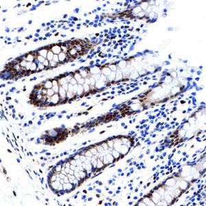
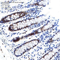
| IHC analysis of NF-κB p65(GB15997).
Sample: Human colon, 4% PFA (G1101) 12-24h.
Antigen retrieval: Citrate buffer (pH 6.0) (G1201), pressure cooker 2 min.
Primary antibody: 1: 2500, 4°C overnight.
Secondary antibody: S-vision poly-HRP conjugated Goat Anti-Rabbit lgG (H+L), Ready to use(G1302), RT, 20min. |
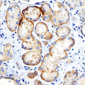
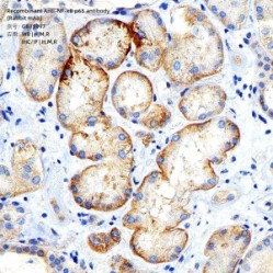
| IHC analysis of NF-κB p65(GB15997).
Sample: Human kidney, 4% PFA (G1101) 12-24h.
Antigen retrieval: Citrate buffer (pH 6.0) (G1201), pressure cooker 2 min.
Primary antibody: 1: 2500, 4°C overnight.
Secondary antibody: S-vision poly-HRP conjugated Goat Anti-Rabbit lgG (H+L), Ready to use(G1302), RT, 20min. |
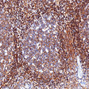
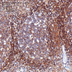
| IHC analysis of NF-κB p65(GB15997).
Sample: Human tonsil, 4% PFA (G1101) 12-24h.
Antigen retrieval: Citrate buffer (pH 6.0) (G1201), pressure cooker 2 min.
Primary antibody: 1: 2500, 4°C overnight.
Secondary antibody: S-vision poly-HRP conjugated Goat Anti-Rabbit lgG (H+L), Ready to use(G1302), RT, 20min. |
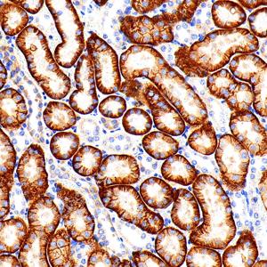
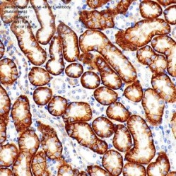
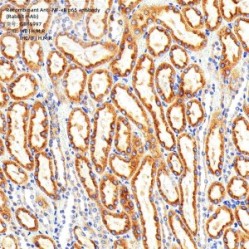
| IHC analysis of NF-κB p65(GB15997).
Sample: Mouse kidney, 4% PFA (G1101) 12-24h.
Antigen retrieval: Citrate buffer (pH 6.0) (G1201), pressure cooker 2 min.
Primary antibody: 1: 2500, 4°C overnight.
Secondary antibody: S-vision poly-HRP conjugated Goat Anti-Rabbit lgG (H+L), Ready to use(G1302), RT, 20min. |
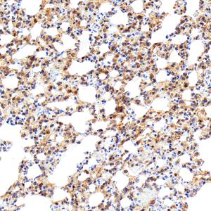
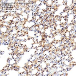
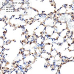
| IHC analysis of NF-κB p65(GB15997).
Sample: Mouse lung injury, 4% PFA (G1101) 12-24h.
Antigen retrieval: Citrate buffer (pH 6.0) (G1201), pressure cooker 2 min.
Primary antibody: 1: 2500, 4°C overnight.
Secondary antibody: S-vision poly-HRP conjugated Goat Anti-Rabbit lgG (H+L), Ready to use(G1302), RT, 20min. |
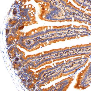
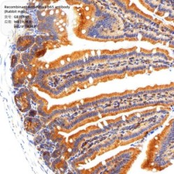
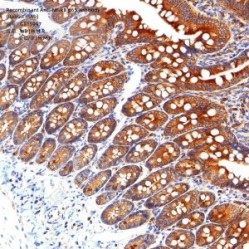
| IHC analysis of NF-κB p65(GB15997).
Sample: Mouse small intestine, 4% PFA (G1101) 12-24h.
Antigen retrieval: Citrate buffer (pH 6.0) (G1201), pressure cooker 2 min.
Primary antibody: 1: 2500, 4°C overnight.
Secondary antibody: S-vision poly-HRP conjugated Goat Anti-Rabbit lgG (H+L), Ready to use(G1302), RT, 20min. |
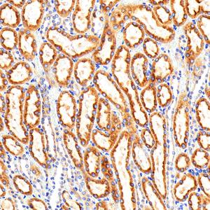
| IHC analysis of NF-κB p65(GB15997).
Sample: Rat kidney, 4% PFA (G1101) 12-24h.
Antigen retrieval: Citrate buffer (pH 6.0) (G1201), pressure cooker 2 min.
Primary antibody: 1: 2500, 4°C overnight.
Secondary antibody: S-vision poly-HRP conjugated Goat Anti-Rabbit lgG (H+L), Ready to use(G1302), RT, 20min. |
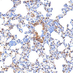
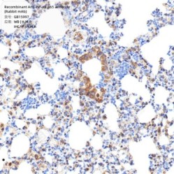
| IHC analysis of NF-κB p65(GB15997).
Sample: Rat lung injury, 4% PFA (G1101) 12-24h.
Antigen retrieval: Citrate buffer (pH 6.0) (G1201), pressure cooker 2 min.
Primary antibody: 1: 2500, 4°C overnight.
Secondary antibody: S-vision poly-HRP conjugated Goat Anti-Rabbit lgG (H+L), Ready to use(G1302), RT, 20min. |
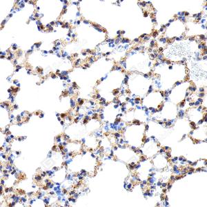
| IHC analysis of NF-κB p65(GB15997).
Sample: Rat lung injury, 4% PFA (G1101) 12-24h.
Antigen retrieval: Citrate buffer (pH 6.0) (G1201), pressure cooker 2 min.
Primary antibody: 1: 2500, 4°C overnight.
Secondary antibody: S-vision poly-HRP conjugated Goat Anti-Rabbit lgG (H+L), Ready to use(G1302), RT, 20min. |
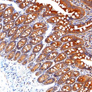
| IHC analysis of NF-κB p65(GB15997).
Sample: Rat small intestine, 4% PFA (G1101) 12-24h.
Antigen retrieval: Citrate buffer (pH 6.0) (G1201), pressure cooker 2 min.
Primary antibody: 1: 2500, 4°C overnight.
Secondary antibody: S-vision poly-HRP conjugated Goat Anti-Rabbit lgG (H+L), Ready to use(G1302), RT, 20min. |
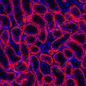
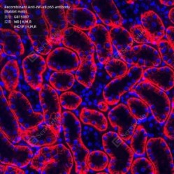
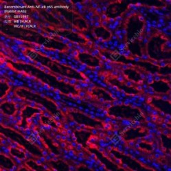
| IF analysis of NF-κB p65(GB15997)red.
Sample: Mouse kindey, 4% PFA (G1101) 12-24h.
Antigen retrieval: Citrate buffer (pH 6.0) (G1201), pressure cooker 2 min.
Blocking buffer: 3% BSA (GC305010) in PBS, RT, 30min.
Primary antibody: 1: 2500, 4°C overnight.
Secondary antibody: Cy3 conjugated Goat Anti-Rabbit IgG (H+L) (GB21303), 1: 300, RT, 1h. |
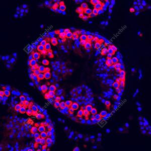
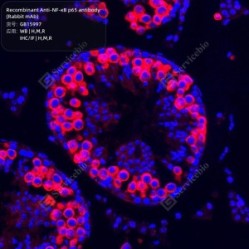
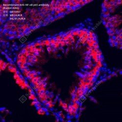
| IF analysis of NF-κB p65(GB15997)red.
Sample: Mouse testis, 4% PFA (G1101) 12-24h.
Antigen retrieval: Citrate buffer (pH 6.0) (G1201), pressure cooker 2 min.
Blocking buffer: 3% BSA (GC305010) in PBS, RT, 30min.
Primary antibody: 1: 2500, 4°C overnight.
Secondary antibody: Cy3 conjugated Goat Anti-Rabbit IgG (H+L) (GB21303), 1: 300, RT, 1h. |
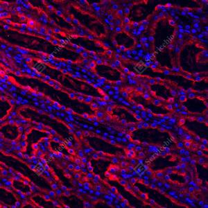
| IF analysis of NF-κB p65(GB15997)red.
Sample: Rat kindey, 4% PFA (G1101) 12-24h.
Antigen retrieval: Citrate buffer (pH 6.0) (G1201), pressure cooker 2 min.
Blocking buffer: 3% BSA (GC305010) in PBS, RT, 30min.
Primary antibody: 1: 2500, 4°C overnight.
Secondary antibody: Cy3 conjugated Goat Anti-Rabbit IgG (H+L) (GB21303), 1: 300, RT, 1h. |
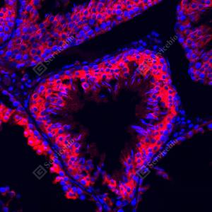
| IF analysis of NF-κB p65(GB15997)red.
Sample: Rat testis, 4% PFA (G1101) 12-24h.
Antigen retrieval: Citrate buffer (pH 6.0) (G1201), pressure cooker 2 min.
Blocking buffer: 3% BSA (GC305010) in PBS, RT, 30min.
Primary antibody: 1: 2500, 4°C overnight.
Secondary antibody: Cy3 conjugated Goat Anti-Rabbit IgG (H+L) (GB21303), 1: 300, RT, 1h. |
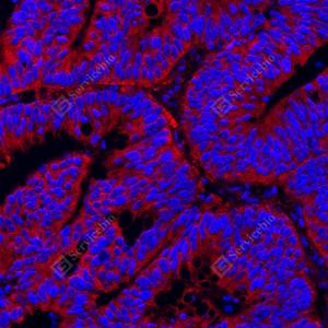
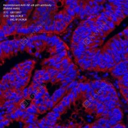
| IF analysis of NF-κB p65(GB15997)red.
Sample: Human colon cancer, 4% PFA (G1101) 12-24h.
Antigen retrieval: Citrate buffer (pH 6.0) (G1201), pressure cooker 2 min.
Blocking buffer: 3% BSA (GC305010) in PBS, RT, 30min.
Primary antibody: 1: 1000, 4°C overnight.
Secondary antibody: Cy3 conjugated Goat Anti-Rabbit IgG (H+L) (GB21303), 1: 300, RT, 1h. |

 Français
 English