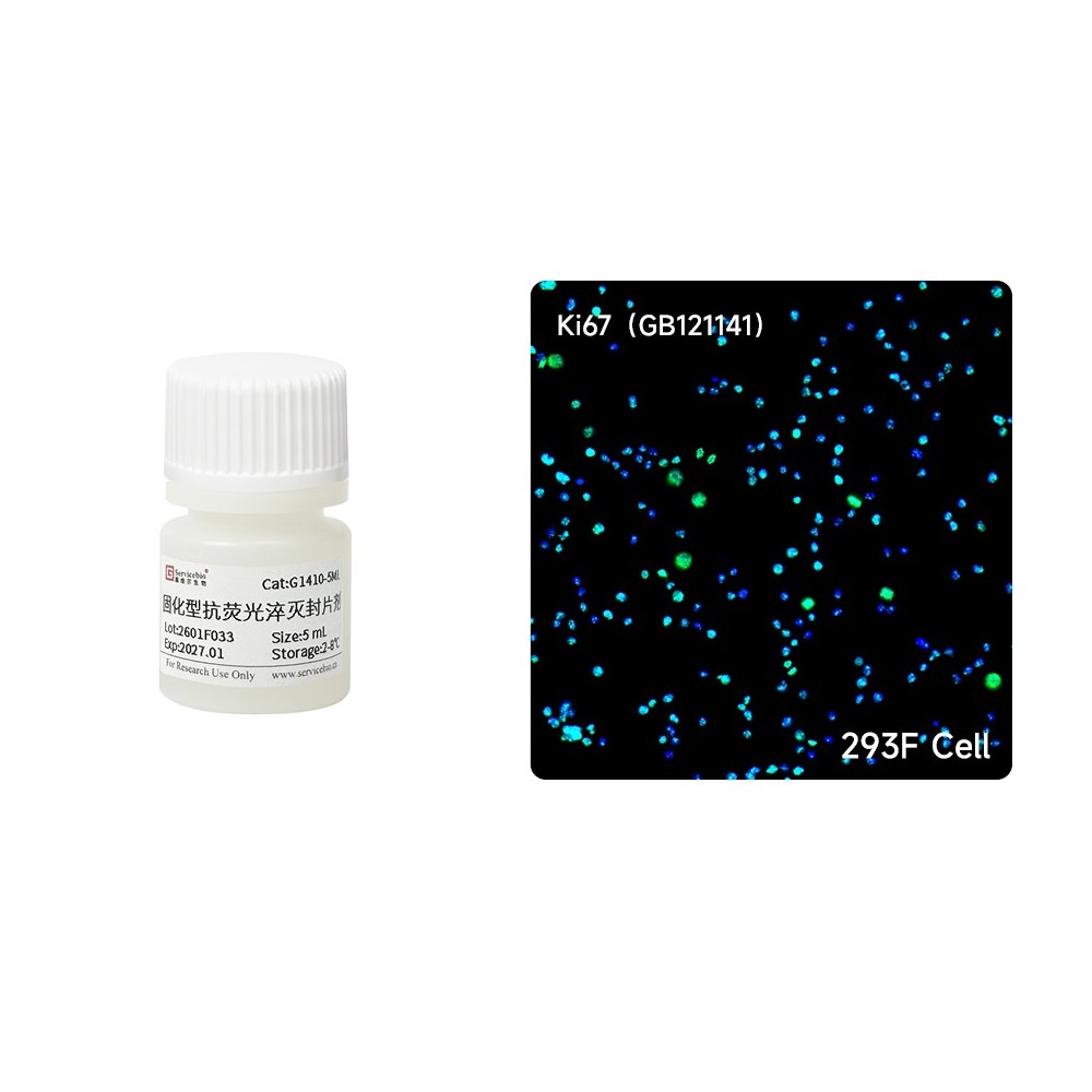
Hardset Anti-fade Mounting Medium
AO-03-G1410-5ML
Product Information
|
Product Name |
Cat. No. |
Spec. |
|
Hardset Anti-fade Mounting Medium |
G1410-5ML |
5 mL |
|
G1410-25ML |
25 mL |
Product Description / Introduction
After fluorescent protein labeling or staining with fluorescent dyes, it is necessary to mount the sample using an aqueous anti-fade mounting medium. Commonly used anti-fade mounting media are primarily composed of glycerol; however, one drawback of these media is that the cover glass may slide after mounting. Although sealing the edges with nail polish can prevent the cover glass from sliding, it introduces other issues such as toxicity, difficulty in removing the cover glass, and difficulty in cleaning up overflowed nail polish.
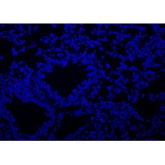
This newly developed mounting medium is specifically designed for sample mounting under aqueous conditions after fluorescent labeling. It is non-toxic and has low viscosity, making it less likely to produce bubbles during mounting. It also has the advantage of rapid curing—after mounting, the slide can be left at room temperature for 30 minutes or more, and the cover glass will no longer slide. Additionally, the cover glass can be easily removed by simply soaking the sample in an aqueous buffer solution.
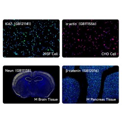
Using this hardset anti-fade mounting medium not only significantly slows down the photobleaching of various common fluorescent dyes and enhances their photostability, but also allows for long-term sample preservation and observation.
Storage and Shipping Conditions
Wet ice transportation, 2-8℃ storage, 12 months shelf life.
Assay Protocol / Procedures
1. After staining, cell or tissue sections must be thoroughly rinsed with PBS or distilled water to remove residual dyes.
2. Gently flick slides to remove excess liquid. Using a Pasteur pipette, dispense 2-3 drops of hardset anti-fade mounting medium onto the sample area. Touch one edge of the cover glass to the mounting medium. Gradually lower the entire cover glass to ensure full contact, minimizing bubble formation.
3. Place slides horizontally in a light-proof container (e.g., drying rack or anhydrous chamber). Incubate at room temperature (≈25°C) for ≥30 minutes until cover glass exhibit no lateral movement. Proceed with fluorescence observation after curing completion.
Note
1. Store the mounting medium undisturbed to prevent bubble formation caused by repeated agitation. Before use, allow the reagent to rewarming at room temperature for 30 minutes. Do not shake or invert the bottle to avoid introducing bubbles.
2. Throughout the dispensing process, air must be prevented from entering the pipette to avoid introducing numerous bubbles into the mounting medium.
3. Recommended Mounting Medium Volume:Apply 2-3 drops of mounting medium per glass slide (75 mm × 25 mm). Over-application causes overflow, adhering slides to wet/chamber boxes and compromising slide retrieval. Inadequate coverage leads to bubble formation and incomplete sample encapsulation.
4. The mounting medium exhibits rapid curing properties. After application on glass slides, cover glass must be placed immediately to prevent premature solidification that would render mounting impossible. Higher temperatures and air circulation accelerate curing speed.At ambient conditions, the medium sets within approximately 10 minutes, beyond which mounting becomes unfeasible.
5. Do not press the cover glass before the mounting medium cures, as this may cause overflow and fingerprint contamination on the cover glass. If overflow occurs after mounting, avoid immediate wiping. Wait until fully cured into a film, then directly peel off excess material.
6. After mounting, place slides horizontally on drying racks (e.g., WGSPB0002) or Waterless humidity chambers (e.g., SIB-20F/SIB-20U). Cure at room temperature (≈25°C) under light-protected conditions for ≥30 minutes until cover glass no longer slide. To expedite curing, apply forced airflow. Do not use ovens for drying, as this induces bubble formation in tissue samples.
7. Immerse the slide directly in aqueous solutions to remove the cover glass if the mounting medium is not fully cured. Gently pry open the edges of the cover glass with a blade, avoiding contact with tissue sections. Submerge the slide in aqueous solutions to complete disassembly.
8. If the slide is dirty and the mounting medium is not fully cured, gently press one corner of the cover glass and wipe the cover glass surface lightly to remove debris. If bubbles are observed during microscopy, apply gentle pressure on the cover glass over the tissue area before complete curing. Displace bubbles toward sample-free areas (e.g., slide edges) without disturbing the tissue.
9. Cured slides may be stored at room temperature (20-25°C) in light-protected conditions for limited duration. For extended preservation, store at 2-8°C with strict light protection.
10. Wear lab coats and disposable gloves during all handling procedures to prevent contamination and chemical exposure
For Research Use Only!
Use collapsible tabs for more detailed information that will help customers make a purchasing decision.
Ex: Shipping and return policies, size guides, and other common questions.